/https://tf-cmsv2-smithsonianmag-media.s3.amazonaws.com/filer/bf/66/bf66ca78-bb89-4f10-a694-e288014e00ca/2020_zebrafish.jpg)
Beauty is in the eye of the beholder, and sometimes capturing beautiful images requires a closer look. For 46 years, Nikon has hosted the Small World photomicrography competition, celebrating photographers who use optical microscopes. At the lens of a microscope, these close-up views reveal intricate patterns and illuminate scientific discoveries.
This year, judges combed through over 2,000 entries from 90 countries, Alan Taylor reports for the Atlantic. And on Tuesday, the company announced the 88 best entries, with 20 receiving top honors.
A stunning photograph of a zebrafish’s head, fins and back took home the first prize. The fish isn’t just a pretty face—it’s also part of a scientific advance. The image captures the zebrafish’s bones and scales in fluorescent blue and its lymphatic vessels in orange. The vessels shown in orange are responsible for clearing toxins and waste from the body, but researchers used to think that only mammals have such systems near their brains. The award-winning photograph shows otherwise.
Zebrafish are far easier to work with in the lab than mammals, and the photograph suggests that scientists can begin to use zebrafish to study the role of the lymphatic system in neurological diseases.
Developmental biologist Daniel Castranova at the National Institutes of Health captured the photograph on a busy day. He used a confocal microscope that captures over 300 individual images that are then stitched together to show the zebrafish at four-times scale in sharp detail. The photograph was one of the last that Castranova captured that day, as he was about to be kicked off of the equipment, he tells Oliver Whang of National Geographic. Then it took days before he saw the results.
“I never even looked at the picture for a couple of weeks,” Castranova tells Science News’ Erin Garcia de Jesus. “And then when I looked at it at some point post-data processing, I was like ‘Wow.’”
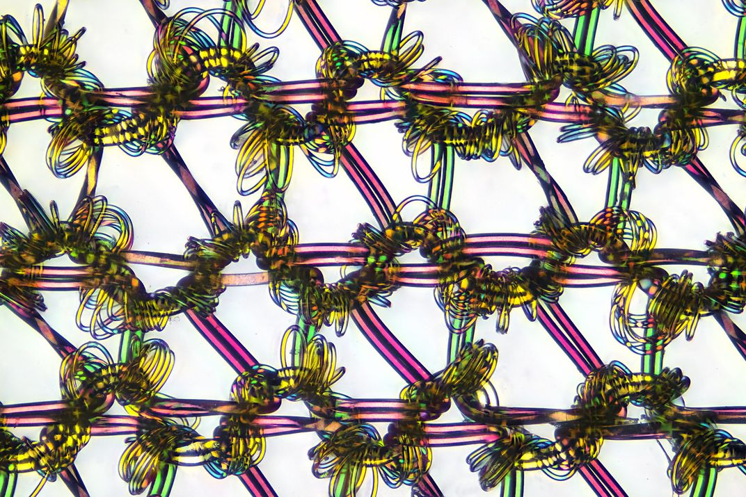
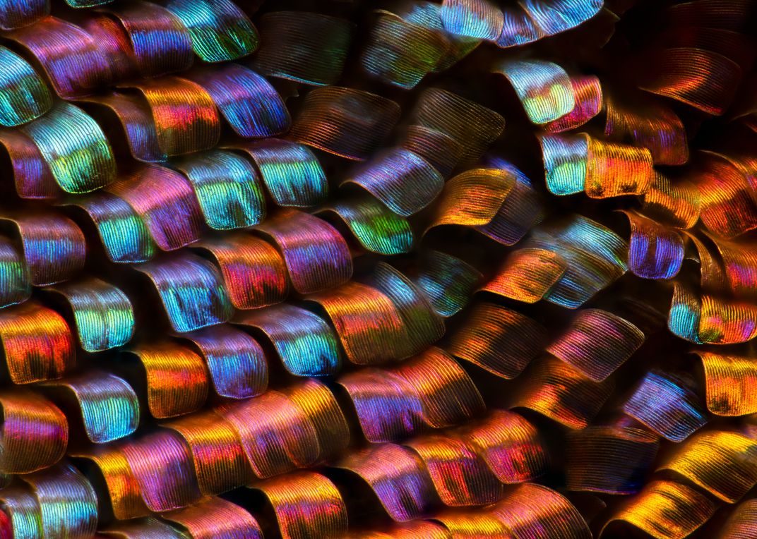
Many of the photographs capture close-up views of biological phenomena, which makes a colorful photograph of nylon stockings stands out. Shot at nine-times magnification, the photograph shows the springy polymers wrapped around red and green straight threads that have been woven together like a chain-link fence.
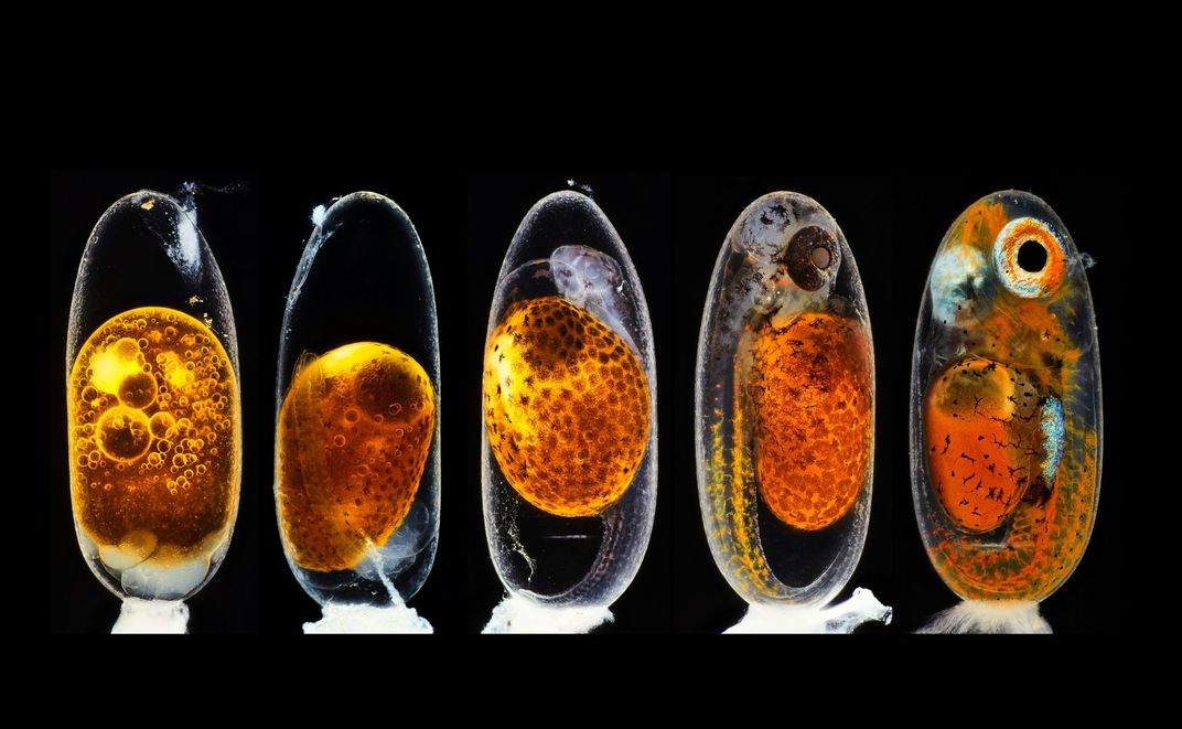
And a series of five photographs of clown fish embryos, which took second place in this year’s competition, show what Nemo looked like from day one to day nine of development. The first embryo in the line-up was photographed hours after conception, and a pack of sperm cells is still visible at the top of the egg. The following embryos show the morning and evening of the third day after fertilization, the fifth day, and the ninth day, shortly before the egg hatched. Capturing the images took special skill because the embryo constantly moved in the egg.
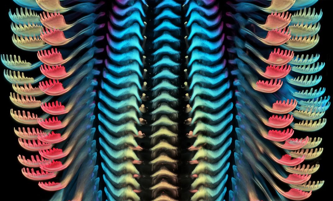
The third-place photograph shows a pastel-hued view of a freshwater snail’s tongue, with its comb-like protrusions flanking the frame. Igor Siwanowicz, a research scientist at the Howard Hughes Medical Institute’s Janelia Research Campus, magnified the snail tongue 40 times and photographed the layers with a laser at different focal lengths. The features that are furthest away from the camera are blue, while the closest are hot pink.
“I chose this image to show that in nature, beauty can be found in the most unexpected places, like a snail’s mouth,” Siwanowicz tells Science News.
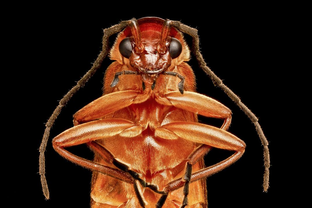
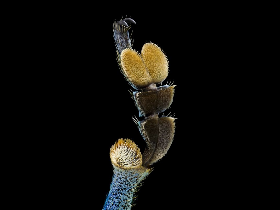
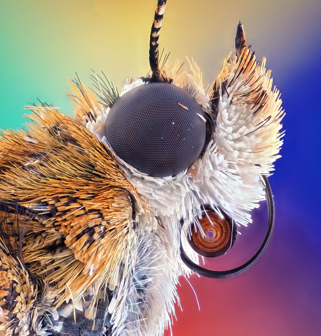
And it wouldn’t be a close-up photography competition without an array of many-legged arthropods. Spiders, beetles, fleas and butterflies took center stage this year, from the fifth-place profile of a bogong moth to a stern-looking portrait of a red soldier beetle. A photograph of a beetle leg shows off its setae, tiny hairs on the exoskeleton that help the beetle perceive touch and sound.
“We are proud to showcase imagery that this blend of research, creativity, imaging technology and expertise can bring to scientific discovery,” Nikon says in a statement. “This year’s first place winner is a stunning example.”