Science Images that Border on Art
This year’s Wellcome Image Award winners pull at your “art” strings. The curious seek out the science behind them
![]()

A false-colored scanning electron micrograph (SEM) of caffeine crystals. “It is a bright, intricate image of something that most of us experience every day,” said James Cutmore, a picture editor at BBC Focus Magazine and a judge for the Wellcome Image Awards. Image by Annie Cavanagh, Wellcome Images.
So many images created in the name of science are brilliant works of art. Magnetic resonance imaging, for instance, produces beautiful reconstructions of the human brain, with all its neural tracts traced in different colors. And, when a geologist photographs a thin slice of peridotite, lit with polarized light, the sample resembles brightly-colored stained glass.
This idea of scientists seeing the artistry in their work certainly hasn’t been lost on Wellcome Images, the world’s leading collection of photographs, X-rays and illustrations chronicling the history of medicine. Each year, the Wellcome Image Awards celebrate the cream of the archive’s new crop of pictures, chosen, as Catherine Draycott, head of Wellcome Images, says, “for their scientific and technical merit as much as for their aesthetic appeal.”
This year’s batch of 16 winners, on display at the Wellcome Collection in London through December 31, depicts cancer cells, bacteria, the connective tissue from a person’s knee and even the surface of a living human’s brain.
“They offer people a chance to get closer to science and research and see it in a different way, as a source of beauty as well as providing important information about ourselves and the world around us,” added Draycott, in a press release.
Here is a sampling, with some scientific explanation to help identify what exactly it is that you are seeing.
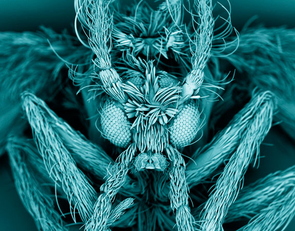
Kevin MacKenzie, who manages a microscopy facility at the University of Aberdeen in Scotland, found a moth fly on his kitchen wall. He decided to take a closer look at the fly under a scanning electron microscope and, in doing so, produced this somewhat menacing image (above). Moth flies, commonly referred to as drain flies, deposit their larvae in sink and bath drains. The flies grow and emerge from the drain, as this one likely did, when they reach adulthood. From anterior to posterior, this fly measures only four to five millimeters. But, under intense magnification, one can see the tiny hairs that cover the insect’s body.

A fluorescence micrograph of a chicken embryo’s vascular system. Image by Vincent Pasque, University of Cambridge, Wellcome Images.
To create this image of a chicken embryo, Vincent Pasque, now at the University of California, Los Angeles, cracked open part of an egg’s shell so that he could inject a fluorescent dye into the embryo’s vascular system. The embryo’s heart pumped the dye throughout the veins and arteries connecting it to the yolk sac. In the center, you can see the embryo’s brain, heart and slender body.
Here, thanks to time-lapse photography, one can see how a cancer cell undergoes mitosis, or cell division, over the course of 16 hours. The red blobs are the DNA in the HeLa cells, and the bright blue represents the cell membranes.
This photograph of the surface of a human brain (selected as the grand prize winner) captures the intimate view that a neurosurgeon had while operating on an epileptic patient. “The arteries are bright scarlet with oxygenated blood, the veins deep purple and the ‘grey matter’ of the brain a flushed, delicate pink,” said Alice Roberts, an anatomist and one of the judges, in a press release. “It is quite extraordinary.”
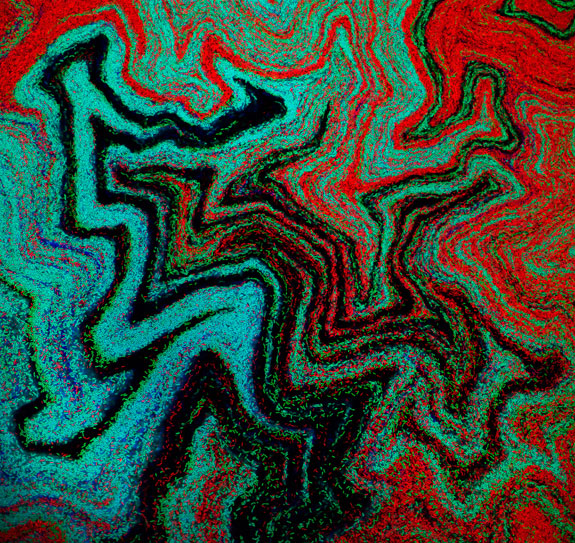
While this may look like a Pointillist painting, it is actually a colony of bacteria growing on a petri dish. Each dot is an individual bacterium, called Bacillus subtilis, and the red, lime green and royal and sky blues represent different lineages. The researchers mixed all of the colors together, at first, but, as the bacteria grew, they reconfigured themselves into mathematically predictable patterns.
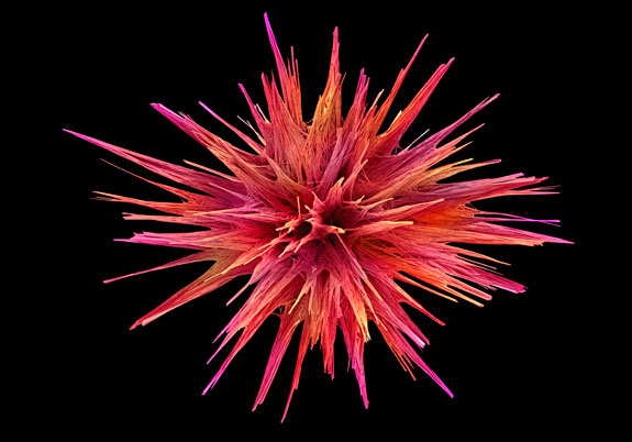
The mesmerizing beauty of this pink, spiky, bur-looking thing might be tainted when you hear the story behind it. What you are looking at is loperamide, a drug used to treat diarrhea. Loperamide targets the nerve fibers in the large intestine, slowing down the movement of the intestine and thereby the food passing through it. As a result, there is more time for some of the water in the food to be reabsorbed into the body.
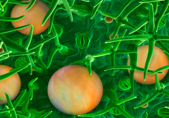
This is a close-up of a lavender leaf, enhanced with bright colors. The green spikes are hair-like growths on the surface of the leaf, called non-glandular trichomes; the orange spheres are glandular trichomes, which contain the shrub’s fragrant oil.
/https://tf-cmsv2-smithsonianmag-media.s3.amazonaws.com/accounts/headshot/megan.png)


/https://tf-cmsv2-smithsonianmag-media.s3.amazonaws.com/accounts/headshot/megan.png)