Watch Corals in Action With New Underwater Microscope
The Benthic Underwater Microscope opens up a whole new age of ocean exploration
/https://tf-cmsv2-smithsonianmag-media.s3.amazonaws.com/filer/00/ea/00ea2f97-558a-4b6b-a7eb-99f7479124eb/polyp.jpg)
Since the discovery of the microscope over 350 years ago, scientists have gotten really good at looking at tiny things, right down to their atoms. But even the most advanced microscopes have one big flaw: they don’t work underwater.
Ocean researchers typically have to collect samples from the briny blue and bring them back to their laboratories to take a good look, which means removing microscopic sea creatures from their habitat, often altering their behavior. But a team of oceanographers recently cracked the problem, developing a Benthic Underwater Microscope that allows a scuba diver to look at and record the tiniest bits of sea life.
“This is important as there are thousands of different millimeter-sized underwater creatures we previously couldn’t study unless they were removed and brought to the lab,” the team writes for The Conversation.
The scope was developed at the Jaffe Lab for Underwater Imaging at the Scripps Oceanographic Institute. It has two parts: a small computer and an imaging unit. The diver uses the computer to control the microscope and camera. And the imaging unit is equipped with a high-powered lens lit by a ring of LED lights connected to a flexible, tunable lens that works kind of like the human eye. It allows the unit to focus on objects only one micron across, about 1/100th the size of a human hair.
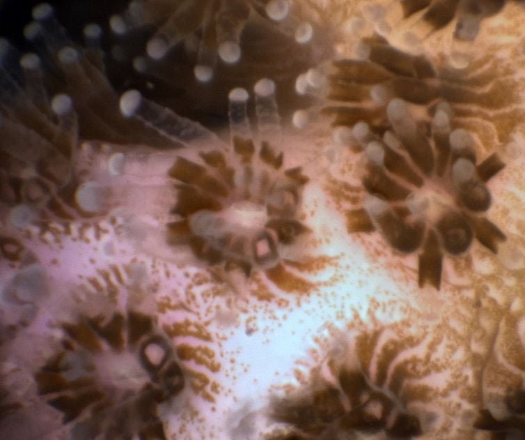
In preliminary runs, the lens has already proven to be a game changer. While testing it at coral reefs in the Red Sea, the team peered at coral polyps, observing never before seen behaviors. Polyps of the same species would occasionally use their tentacles to embrace their neighbors, “potentially to share food, in what we called polyp kissing,” the team writes at The Conversation.

They also noticed that when polyps of different species were placed close to each other, they attacked. The stronger polyp would send out filaments, which are essentially part of its gut, covering the nearby polyp in digestive enzymes, reports Megan Daley at the Los Angeles Times.
“They use those filaments to basically digest the neighbor next to them,” says Andrew Mullen, the grad student who helped develop the microscope and lead author of a study on the microscope in the journal Nature Communications. It took one polyp almost all night to digest its opponent.
In Maui, the researchers used the system to examine coral bleaching and how algae colonizes and eventually smothers damaged coral reefs. They discovered a unique honeycomb pattern that the algae follows when colonizing the reef, something not seen before in the lab.
When oceanographer Victor Smetacek conceived the idea in 2002, he pondered if an underwater microscope could “do for microbial ecology what Galileo’s telescope did for astronomy.” And these preliminary trials suggest the microscope is well on its way. There are many questions this fancy gadget can now help answer, including how kelp propagate, how coral reef diseases progress and how coral larvae develop.
The system is not widely available, but until it is, the team says it's making its microscope available to the scientific community and will travel to research projects around the world to help take photos and videos.