Prize-Winning Photos Capture the Big Beauty of a Microscopic World
Nikon’s Small World Photography Competition celebrates the gorgeous details of nature
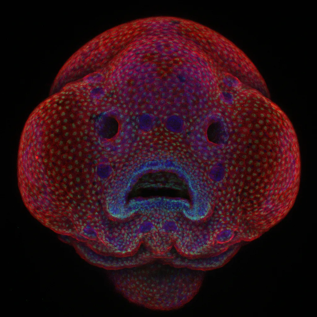
Oscar Ruiz, medical researcher at the University of Texas, studies facial abnormalities by examining cell development in the minute faces of live zebrafish embryos. He takes thousands of images of these microscopic subjects to study their genetics with the hopes of pinning down the genes that control the development of abnormalities such as cleft lips or palates in humans.
This week one of the thousands of images he takes for his work won first place in Nikon’s 42nd annual Small World Photography competition. The winning images were selected out of a pool of over 2,000 submissions by a panel of judges, including two biologists, two science journalists and a high-energy physics researcher.
Imaging live embryo is no easy task. Ruiz commonly captured the fish in profile or from the top, but getting a straight-on image of the fish’s developing face was difficult. So Ruiz experimented with mounting the developing fish in agarose, a type of gelatinous material, and snapped away with his confocal microscope, which uses a laser and software to keep the whole subject in focus.
The method worked, and he was able to create an up-close picture of the developing zebrafish face. “[This image] was the first one we got just the way we wanted,” he says.
The success of Ruiz’s new imaging method actually led him to begin building an image atlas of facial skin cells of the developing zebrafish. Once completed, he and his colleagues will be able to manipulate the fish genes to identify links between the genes and facial cells, which may be applicable to mutations in the human face.
To study the cells, Ruiz uses a stain that causes the nuclei in the cells of the fish to fluoresce, then takes photos and videos over timed intervals to record how those cells move and change. “Basically you start with a little embryo that has no face, then at the end you have a fish that has a face and a mouth and eyes and everything,” he says. Through this research, Ruiz and his team hope to answer fundamental questions about how facial features develop to eventually figure out how to fix these developmental abnormalities.
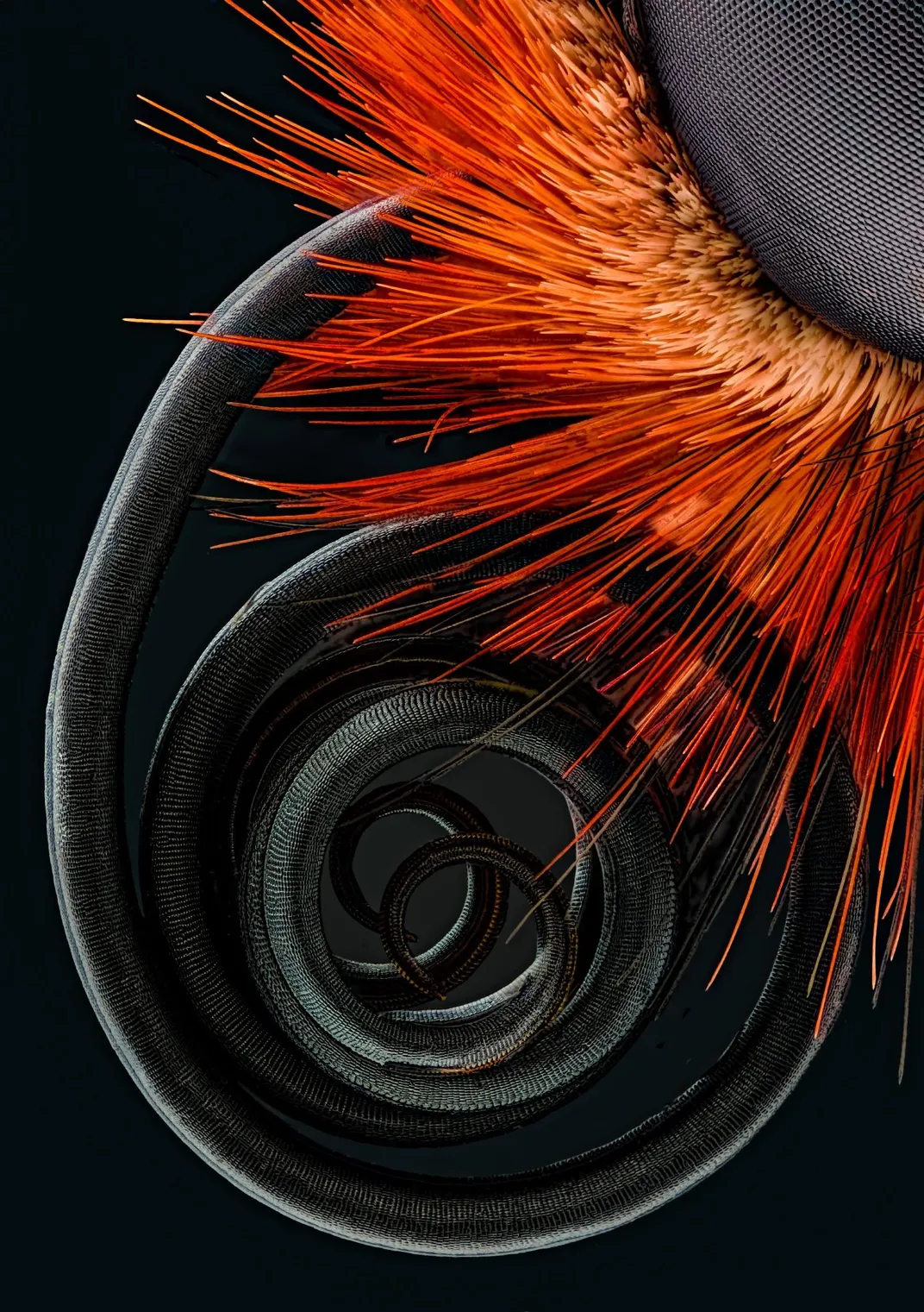
Most of the other images in the final 20 have similarly compelling stories. From glimpses into medical research to gazing into a spider’s eyes, “each image evokes a powerful reaction from our judges,” Nikon's communication manager Eric Flem says in a press release. “Every year we’re looking for that image that makes people lean forward in their seats, sparks their curiosity and leads them to ask new questions.”
Though anyone can enter the contest, it presents an unusual opportunity for researchers in a range of disciplines to show off their work to the general public and help people better understand the research that happens behind closed doors. “As scientists, we work on taxpayer dollars and the general public doesn’t know what we’re researching or see what we’re doing,” says Ruiz. “The more that people see the more they are okay with funding science.”
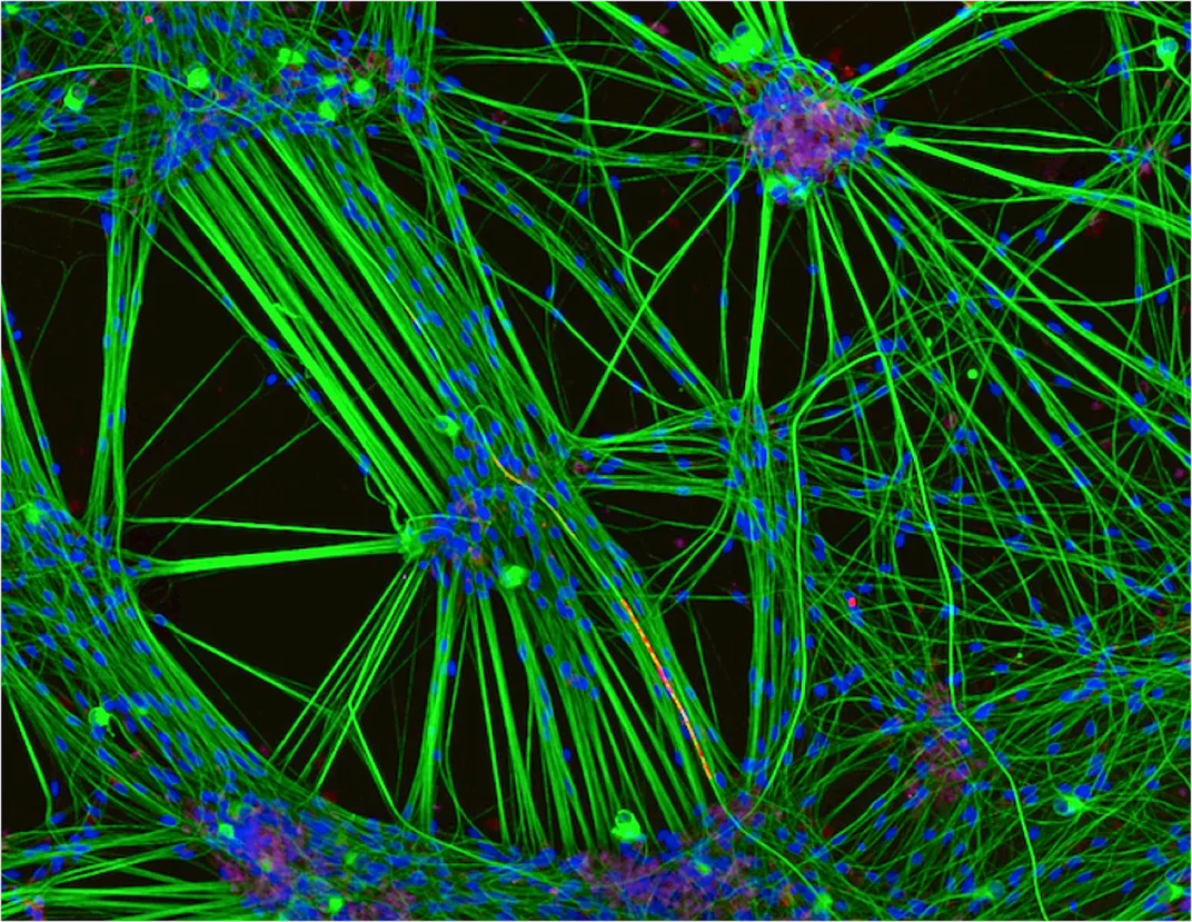
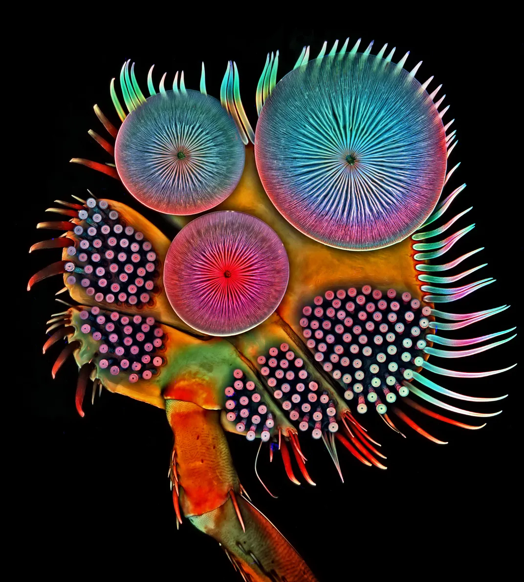
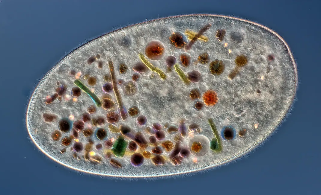
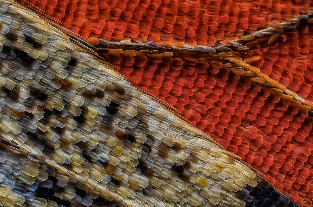
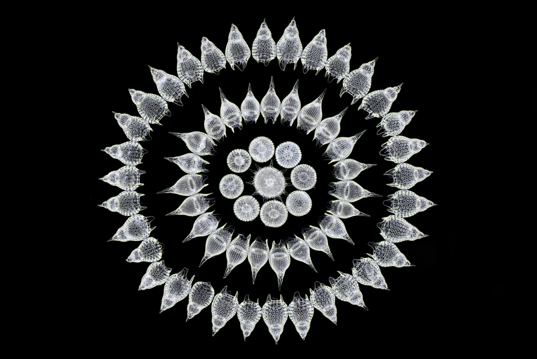
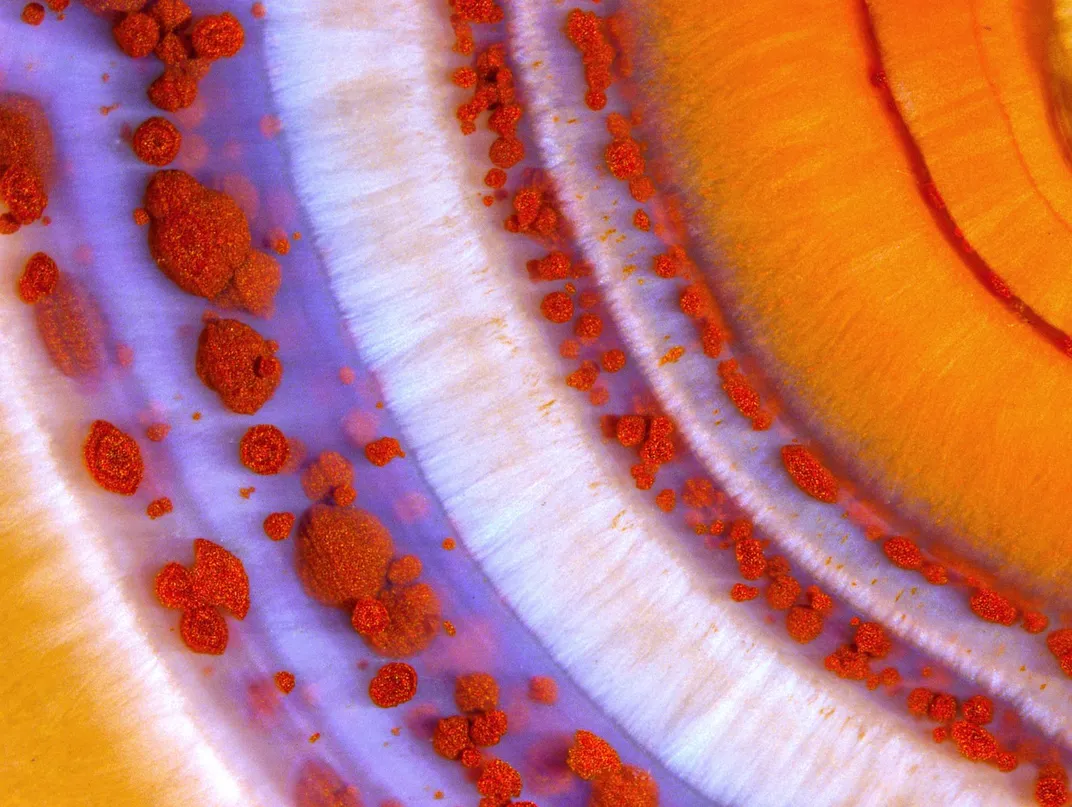
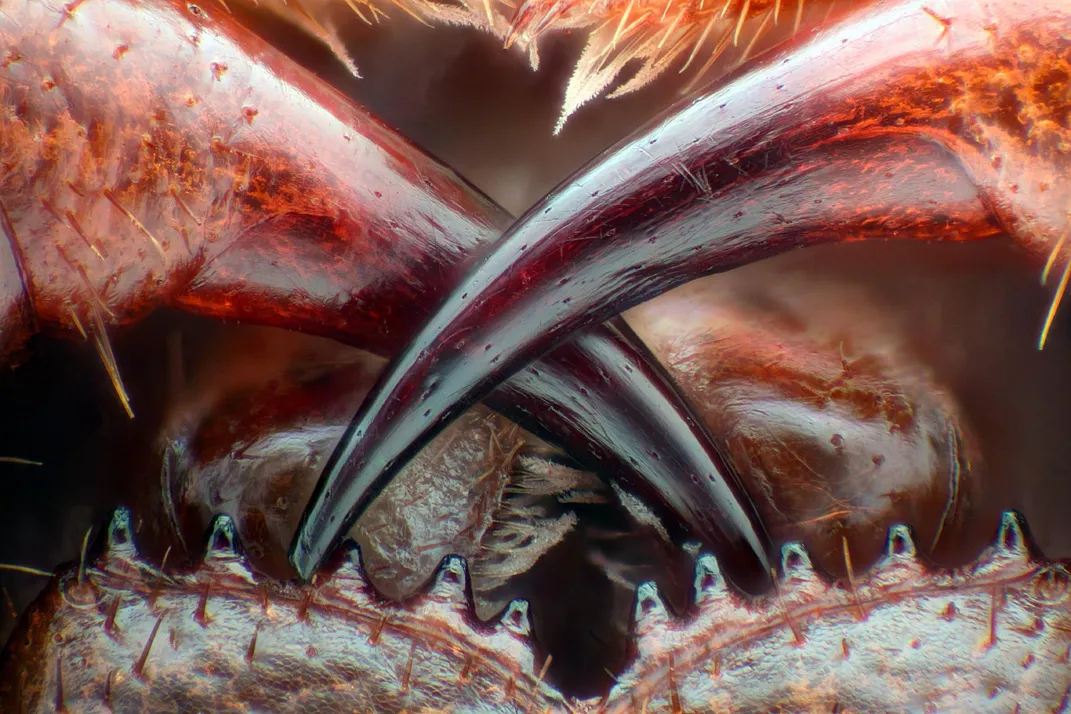
Other images include color pictures of human neurons, close-ups of insect legs and wings, chemical reactions, cell division and microscopic organisms. Some images only slightly magnify their subjects, while others show things that are usually 200 times smaller. The images were taken with a range microscopes, processing and lighting. Some are basic snapshots through a microscope. Others, like Ruiz’s winning shot, use confocal microscopy—a method that captures slices of the object at different depths.
Though the judges have made their decision, public voting on the images will continue until October 25 when a Popular Vote winner will be selected.
