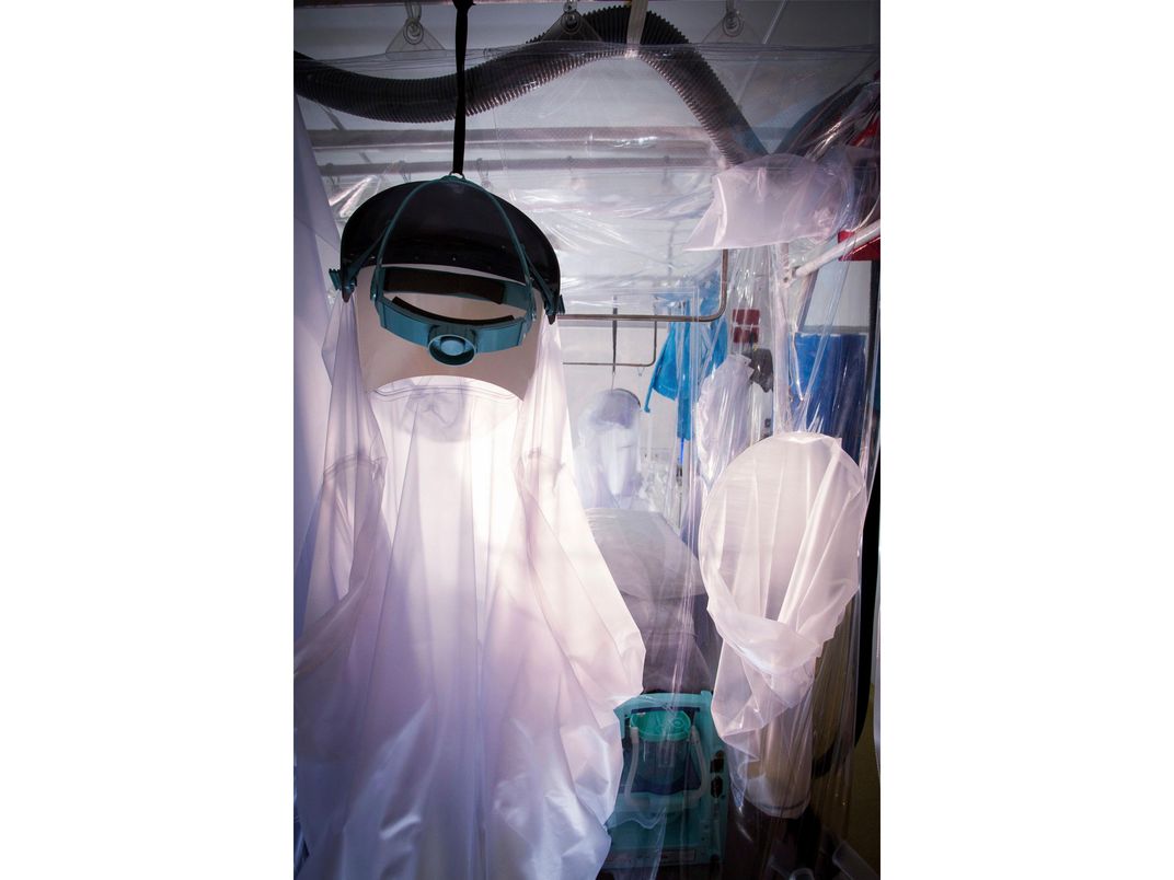
This special see-through tent surrounds a bed in the Royal Free Hospital in London for patients with dangerous infectious diseases, like Ebola. The tent quarantines the patient to allow safe treatment—even the air is cleaned before it is released from the tent.
David Bishop, Royal Free Hospital, London
/
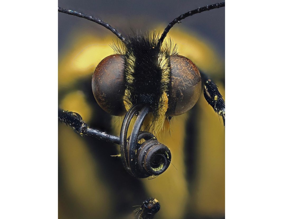
This swallowtail butterfly is ready for its close up. Butterflies have two big round eyes that can detect quick movements and two antennae for sensing their surroundings. The long coil shown here is actually a tube-like 'tongue' that it uses to drink nectar from flowers.
Daniel Saftner, Macroscopic Solutions
/
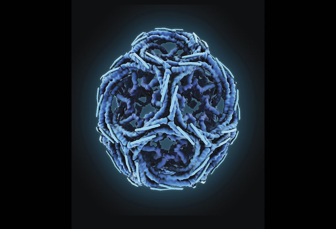
This minute cage-like structure is actually a protein that can help shuttle molecules in and around the cell—and can be broken down when unneeded. Some disease-causing toxins and germs can hijack this process, using it to infect cells.
Maria Voigt, RCSB Protein Data Bank
/
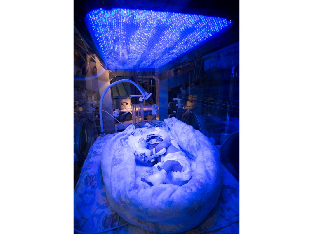
This premature baby has jaundice, a common infant ailment. A preemie's liver often doesn't function well enough to rid the body of bilirubin—a substance naturally produced from the breakdown of blood cells and whose buildup turns the skin and eyes yellow. The baby is being treated under a blue colored light, which can help eliminate the bilirubin.
David Bishop, Royal Free Hospital, London
/
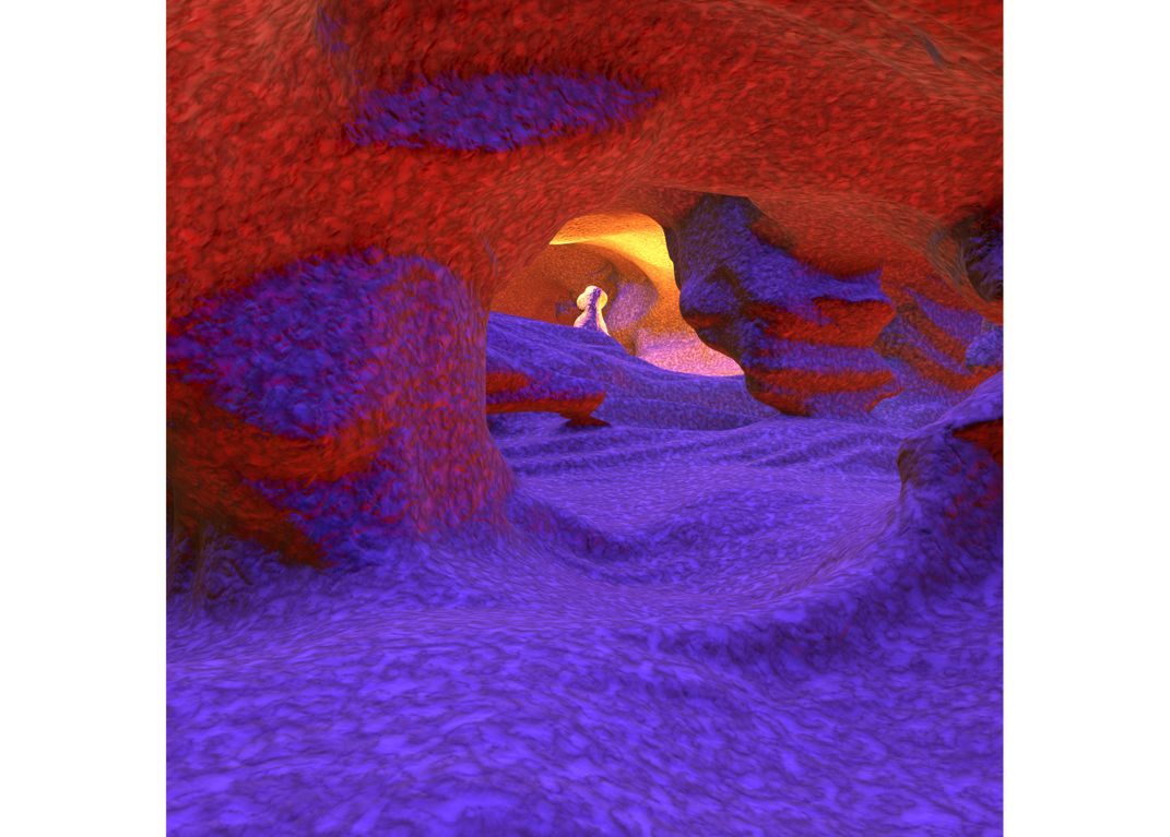
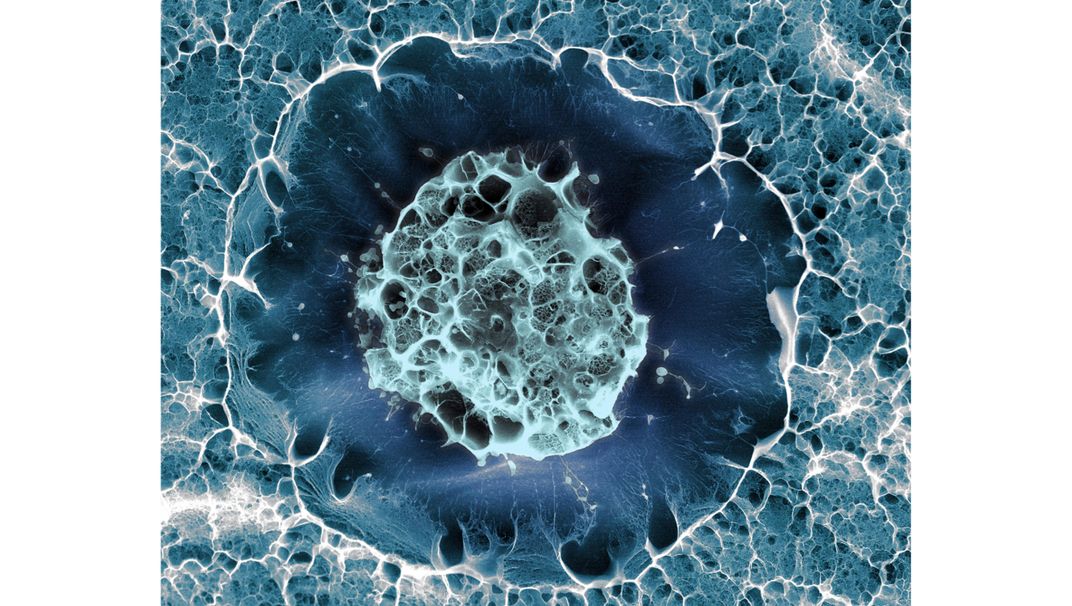
Though this looks like a cavern, this image details the inside of the human eye. Blood flows through these tiny tunnels—each slightly larger than the thickness of an average human hair.
Peter Maloca, University of Basel
/
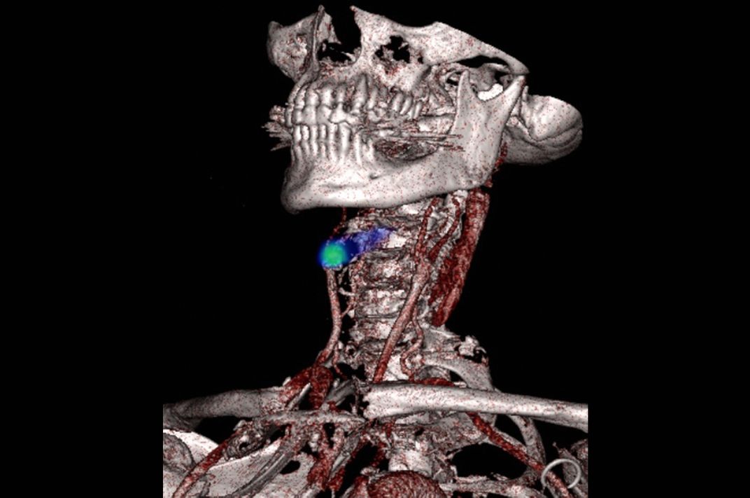
This medical scan shows a blocked blood vessel highlighted in green. The blood vessel in question carries blood to the brain, but this blockage can damage brain function—a process more commonly known as a stroke.
/
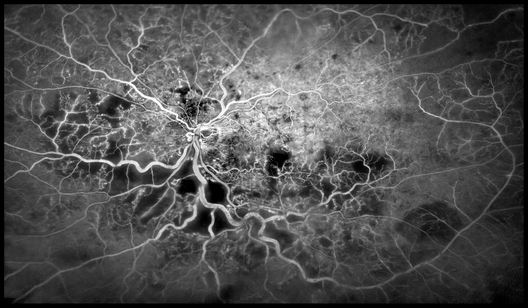
These white spidery lines are the tiny blood vessels inside a human eye. Blood travels through these tubes to keep the eye functioning properly. But if the tubes become blocked or leak, the person's sight will suffer.
Kim Baxter, Cambridge University Hospitals NHS Foundation Trust
/
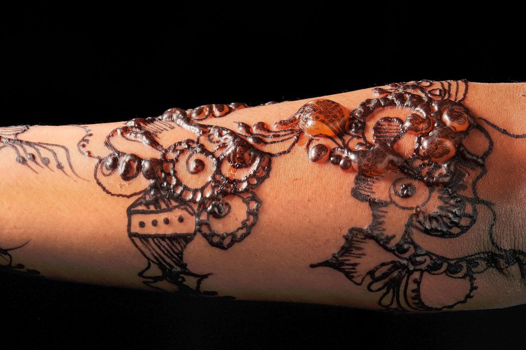
Henna is commonly used to temporarily stain skin or hair orange-brown—an additional chemical turns the color black. But the color comes at a cost. It can cause blistering allergic reactions, as shown here.
Nicola Kelley, Cardiff and Vale University Hospital NHS Trust
/
A close-up look at the scales of a Madagascan sunset moth. Though the moth's bright colors often trick people into calling it a butterfly, the color is actually an illusion. The curved scales bounce away the light, giving the near-colorless wings vivid hues.
Mark R Smith, Macroscopic Solutions
/
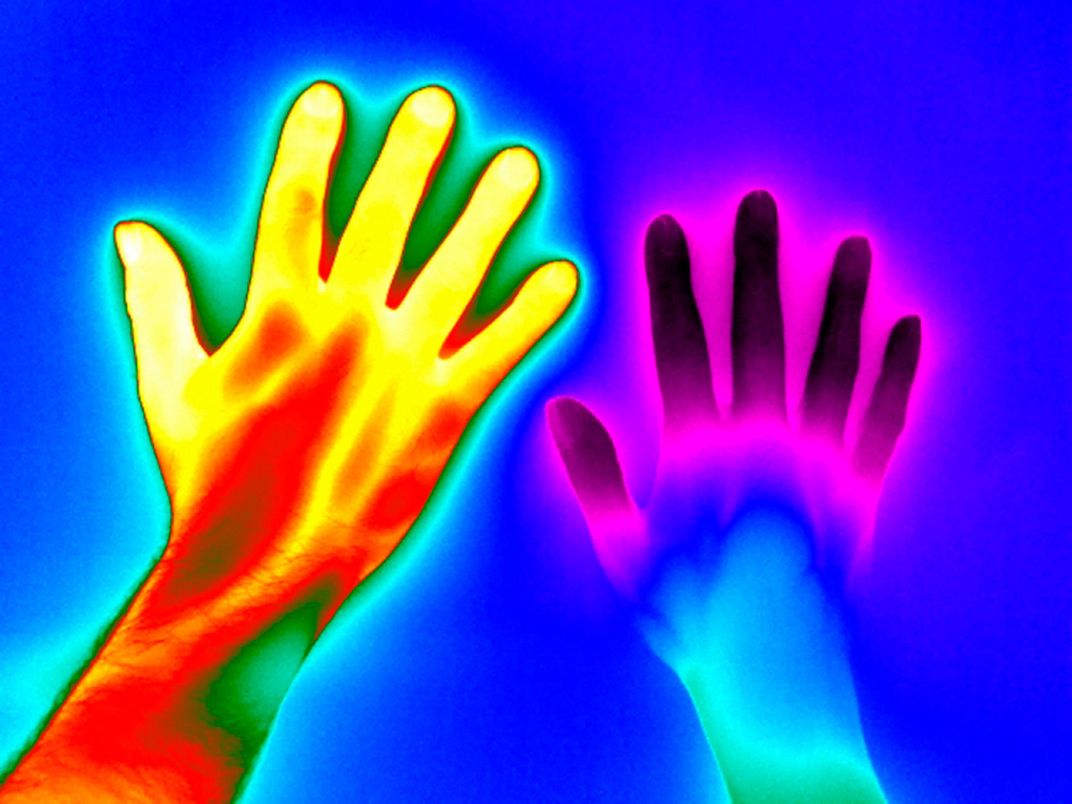
This picture shows the temperature of two people’s hands. The one on the left is from a healthy person while the one on the right is from someone with Raynaud’s disease—an ailment that often causes cold hands and feet.
Matthew Clavey, Thermal Vision Research
/
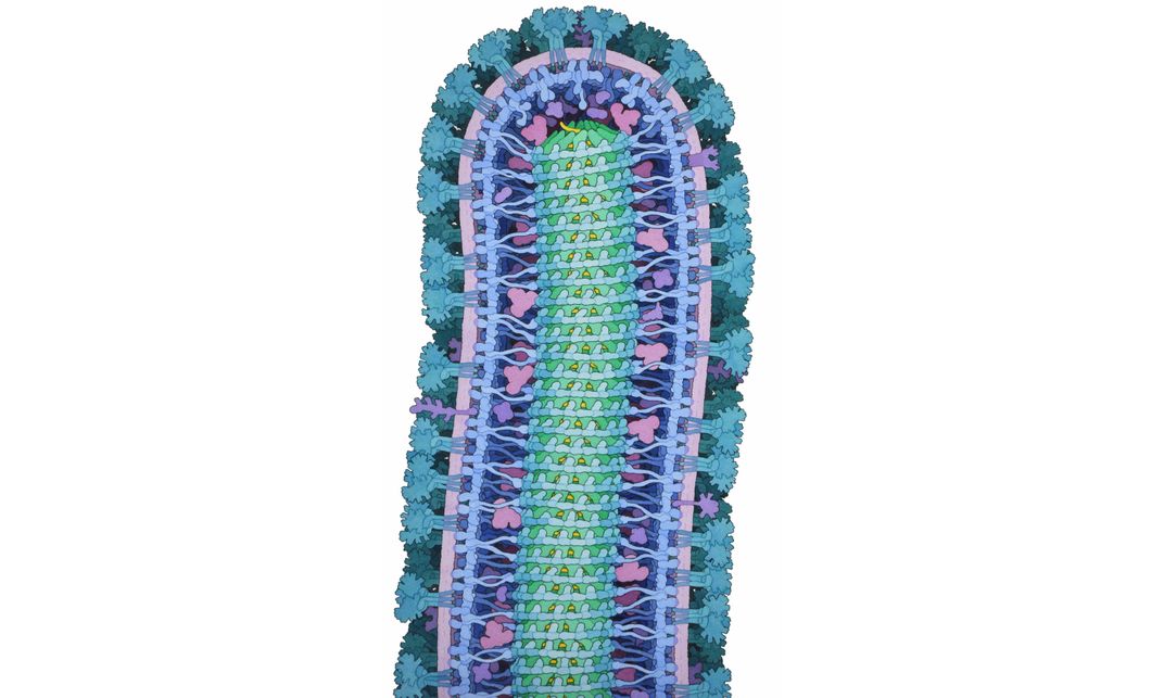
This watercolor and ink illustration details the inner structure of the tiny and destructive Ebola virus. First cropping up in Africa in the mid-1970s, the virus spreads through contact with an infected person's body fluids.
David S Goodsell, RCSB Protein Data Bank
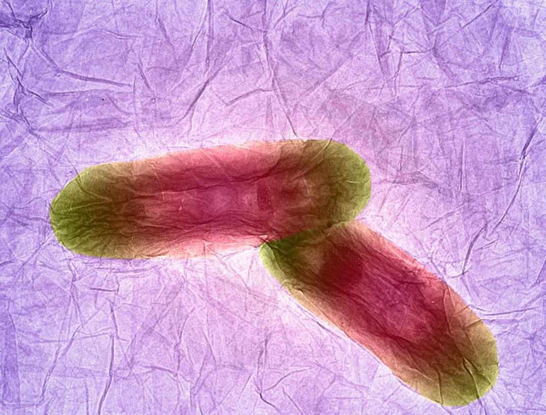
Two bacteria sit on an extremely thin sheet of carbon known as graphene—one of thinnest, strongest materials so far discovered. Though these two bacteria accidentally became entangled in the sheet, researchers are trying to intentionally stick in different medicines to deliver drugs to specific parts of the body.
Izzat Suffian, Kuo-Ching Mei, Houmam Kafa and Khuloud T Al-Jamal, King’s College London
/
This psychedelic form is actually a map of pathways inside a human brain. The different colors show the ways various parts of the brain communicate—left with right sides in red, front with back in green, and the brain to the rest of the body in blue.
Alfred Anwander, Max Planck Institute for Human Cognitive and Brain Sciences
/
Four times the size of a human heart, this preserved cow heart shows the intricate innards of a very important organ.
Michael Frank, Royal Veterinary College
/
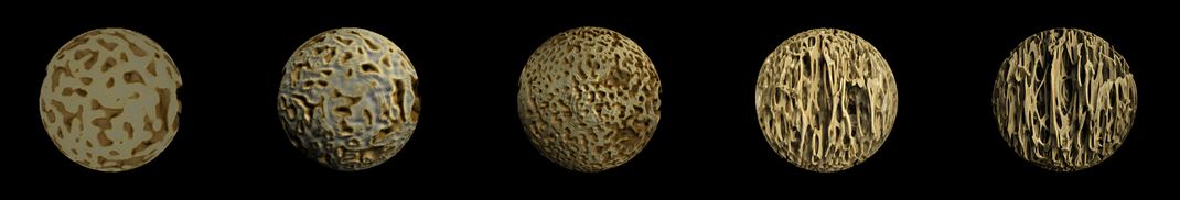
As babies grow, the the textures of their bones develop along with their bodies. This image tracks these changes in 19th century skeletons of children from three months before birth (left) all the way to 2.5 years old (right).
Frank Acquaah
/
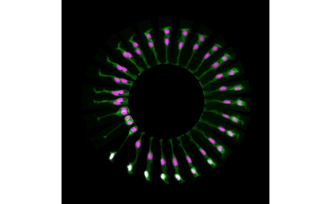
This circle of pictures shows the different stages of a stem cell splitting inside the brain of a zebrafish before it hatches. Starting as a single purple blob at the 8 o’clock position, the cell splits in two, the second blob eventually appearing white.
Paula Alexandre, University College London
/
A close-up look at parasites that cause the disease toxoplasmosis. Sometimes found in cat poop and uncooked meat, the parasites must inhabit another living creature for food and shelter.
Leandro Lemgruber, University of Glasgow
/
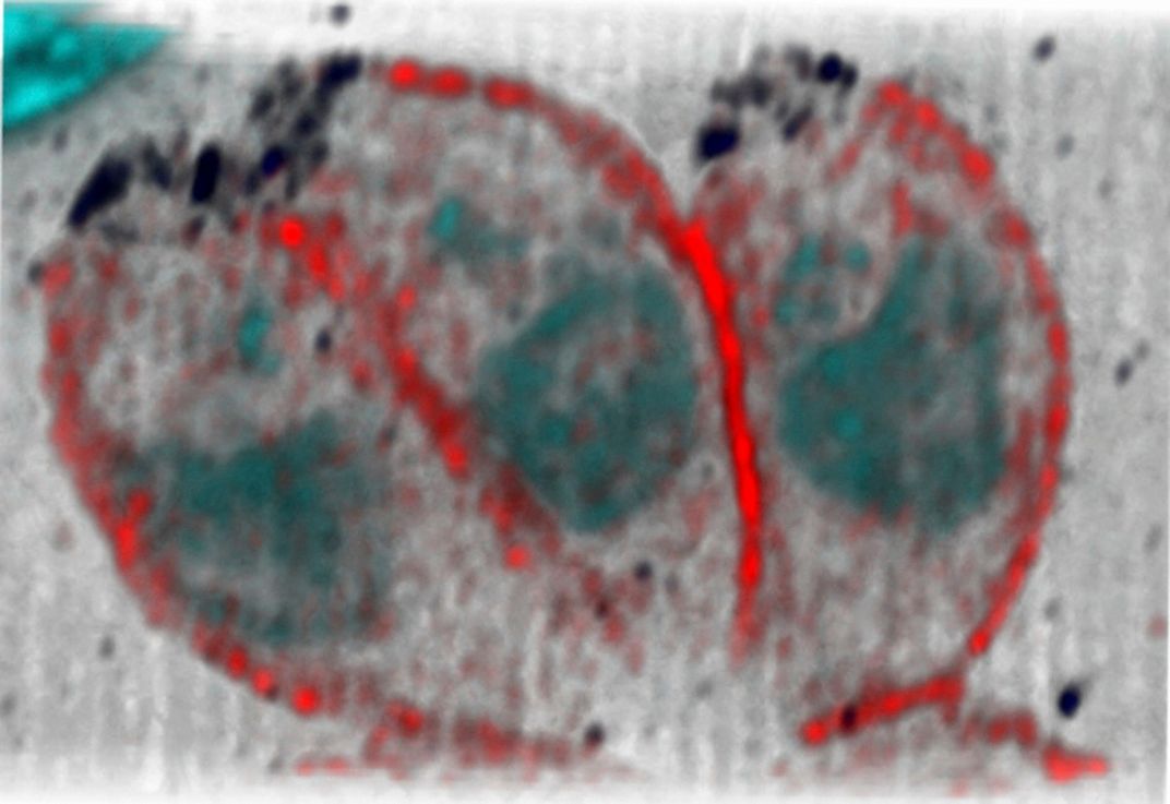
This image captures a single human stem cell, which can diversify as needed to form other types of cells. This particular stem cell came from inside a human hip bone.
Sílvia A Ferreira, Cristina Lopo and Eileen Gentleman, King’s College London
/
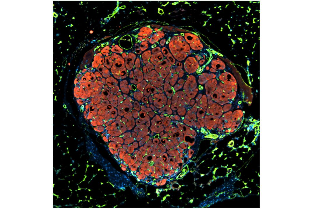
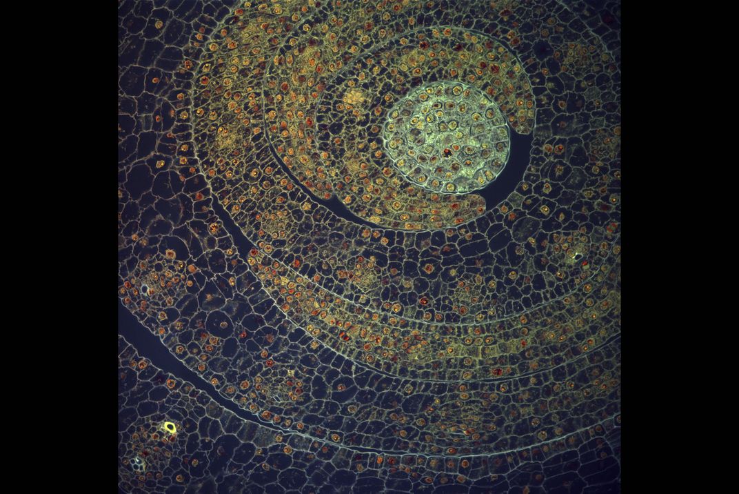
Only spanning about 0.01 inches across, this image gives a peek inside a cluster of curled leaves from a young corn plant. Many box-like cells make up each crescent-shaped leaf cross section. And within each cell is a tiny orange nucleus—its central control center.
Fernán Federici, Pontificia Universidad Católica de Chile and University of Cambridge
What do brains, butterflies, bacteria and blisters have in common? They’re all subjects of this year’s Wellcome Image Awards winners—and show just how emotional and evocative the visual side of science can be.
Every image selected for the 2016 awards shows a different side of medicine and science. The images are collected by Wellcome Images, a medical picture library with a vast collection of scientific imagery, and judged by a panel of science communication and biomedical experts.
The 20 finalists depict everything from moth scales to a premature baby receiving light therapy. One of these images reveals the intricate connections of the human brain—each nerve fiber is color-coded in the composite image. The image is the work of neuroscientist Alfred Anwander, of Germany’s Max Planck Institute, who stitched it together from virtual slices of the brain using diffusion imaging, a type of MRI that tracks the movement and direction of water molecules within the brain.
The awards were established in 1997 in thanks to the contributors of the database for their spectacular imagery. Each year the panel selects finalists and a grand prize winner. This overall winner will be announced for the latest competition at the awards ceremony on March 15.
Since all of the winning images are available under Creative Commons licenses, you can use them any way you want. Even better, you’ll soon be able to view them at science and technology institutions all over the world, including the MIT Koch Institute in Massachusetts, the Africa Center for Population Health in South Africa and the Polytechnic Museum in Moscow. After all, science knows no language—and with pictures like these, it’s easy to understand why.
Get the latest stories in your inbox every weekday.
Erin Blakemore is a Boulder, Colorado-based journalist. Her work has appeared in publications like The Washington Post, TIME, mental_floss, Popular Science and JSTOR Daily. Learn more at erinblakemore.com.
/https://tf-cmsv2-smithsonianmag-media.s3.amazonaws.com/accounts/headshot/erin.png)

/https://tf-cmsv2-smithsonianmag-media.s3.amazonaws.com/filer/a8/dd/a8ddb439-6c3d-44f6-8dc9-547c48c81466/b0010280.jpg)

/https://tf-cmsv2-smithsonianmag-media.s3.amazonaws.com/accounts/headshot/erin.png)