See a Google-Earth-Like View of an Embryo, Down to an Individual Cell
A new technology combines thousands of individual images to create a zoom-able picture of living tissue, down to the cellular level
/https://tf-cmsv2-smithsonianmag-media.s3.amazonaws.com/accounts/headshot/joseph-stromberg-240.jpg)
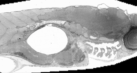
A zebrafish embryo viewed as a whole, composed of more than 26,000 detailed images. Photo via the Journal of Cell Biology
When Google Earth first came out in 2005, many of us had a similar experience. Staring wide-eyed at our computer screen, we zoomed in from an image of Earth in space to a view of North America, then the United States, then our home state, then city, then neighborhood, eventually mesmerized by a view of just our own house or apartment building.

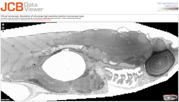
Subsequent zooms on the combined zebrafish embryo image. Photo via the Journal of Cell Biology
Now, a research team from Leiden University Medical Center in the Netherlands has made the same experience possible for a piece of biological tissue. As detailed in an article published yesterday in the Journal of Cell Biology, the researchers have created a new technology they call “virtual nanoscopy.” By stitching together thousands of images from an electron microscope, they allow viewers to zoom from a tissue-level view down to see inside individuals cells in detail. You can experience the technology for yourself on the journal’s website, with a zebrafish embryo image used as a demonstration.
Since the 1950s, electronic microscopes have allowed biologists to see the structures inside cells in remarkable detail. The problem—especially for laypeople—is that these images are so zoomed in it’s hard to tell exactly what you’re looking at. Tiny portions of a cell are captured in each picture, but viewed in isolation, they’re hard to mentally imagine in terms of the whole cell, let alone a piece of tissue or an entire organism.
Additionally, the research process itself suffers from the limitations of this approach. Microscopists typically scan the specimen to produce a lower-resolution overview, and then zoom in to produce detailed images only in the areas that seem to be of interest. Going back later to take close-ups of other areas can often be difficult, if not impossible, the researchers note, because certain types of preserved specimens can deteriorate over time.
In response, the research team developed a new way of combining thousands of distinct electron microscope images to create a coherent and interactive whole. As part of the process, thousands of slightly overlapping images are collected in one initial phase. Then, an automated software program virtually stitches them together, using metadata on the individual images’ orientation and an algorithm that compares similar features in each of them to determine exactly where they should be placed.
The zebrafish embryo shown is composed of more than 26,000 individual images. The enormous file weighs in at a total of 281 gigapixels, with 16 million pixels per inch. The entire embryo is 1.5 mm long, and you can move from a zoomed-out picture of the whole to a detailed view of structures, such as a nucleus, within a specific cell.
The new technology will serve as more than Internet entertainment for the scientifically-inclined. The researchers state that their new method can be used to help other scientists make discoveries, because they will be more able to relate structures with functions on a variety of scales. As evidence, they used the technique to analyze the zebrafish embryo, human skin tissue, a mouse embryo and mouse kidney cells.
/https://tf-cmsv2-smithsonianmag-media.s3.amazonaws.com/accounts/headshot/joseph-stromberg-240.jpg)