A Postmortem of the Most Famous Brain in Neuroscience History
Patient H.M.’s brain has been sliced and digitized, leading to new insights for scientists
/https://tf-cmsv2-smithsonianmag-media.s3.amazonaws.com/accounts/headshot/joseph-stromberg-240.jpg)
/https://tf-cmsv2-smithsonianmag-media.s3.amazonaws.com/filer/fb/49/fb492876-abb1-48a2-be14-b5b5b97784e3/hm_brain_1.jpg)
On August 25, 1953, a 27-year-old Connecticut native named Henry Molaison underwent brain surgery to treat the seizures he chronically suffered from as a result of epilepsy. Hartford Hospital neurosurgeon William Beecher Scoville, who had previously determined the brain regions where Molaison's seizures originated, removed a fist-sized chunk of brain tissue that included parts of both his left and right medial temporal lobes.
When Molaison awoke after the surgery, his epilepsy was largely cured. But removing so much brain tissue—and, in particular, a structure called the hippocampus—led to an entirely new problem for H.M, as he'd soon be called in the scientific literature to protect his privacy.
From that moment on, he was unable to create memories of any new events, names, people, places or experiences. He also lost most of the memories he'd formed in the years leading up to surgery. In the most fundamental sense possible, H.M. lived entirely in the moment.
"At this moment, everything looks clear to me, but what happened just before?" he once said. "That’s what worries me. It’s like waking from a dream. I just don’t remember." Although he interacted with the same nurses and doctors day after day, each time he saw them he had no idea he'd ever met them before. He remained a perfectly intelligent, perceptive person, but was unable to hold down a job or live on his own. Without the connective tissue of long-term memory, his life was reduced to a series of incoherent, isolated moments.
Out of this tragic misfortune came an unintended benefit. For decades, neuroscientists closely studied H.M., making groundbreaking discoveries about memory formation based on his condition. He voluntarily participated in testing almost continually, and by the end, he was widely known as the most important patient in neuroscience history.
When he died in 2008, researchers led by Jacopo Annese of UC San Diego froze his brain in gelatin and cut it into 2,401 ultra-thin slices for further research. Now, in a paper published today in Nature Communications, they've announced the results of their analysis. By using the slices to create a 3D microscopically-detailed model of H.M.'s brain, they've identified a previously-unknown lesion caused by the surgery, a finding that could shed further light on the anatomical structures responsible for memory.
In the decades after H.M.'s surgery, researchers such as Brenda Milner and Suzanne Corkin studied H.M.'s memory limitations and used them to pioneer the nascent field of memory study. With records of the 1953 procedure, they were even able to link particular anatomical areas that H.M. was missing with memory functions.
Previously, many had believed that it was impossible to assign functions to physical stuctures in this way, but H.M.'s unique case opened up new possibilities. He was incapable of storing new information in his explicit memory—the type of memory that allows us to consciously remember experiences and pieces of new information—but could remember pieces of information over a very short time period (up to about 20 seconds), evidence that his short-term memory was somewhat intact. He could also learn and retain new skills, even if he didn't remember the actual act of learning them.
These fine distinctions led scientists to distinguish between procedural memory—the unconscious memory that allows us to perform motor activities, like driving—and explicit memory. Additionally, that H.M. couldn't form new explicit memories but had undamaged childhood memories highlighted the difference between memory encoding and memory retrieval (he could still perform the latter, but not the former). Perhaps most importantly, the fact that he was missing his hippocampus suggested that the structure was crucially involved in the encoding of long-term explicit memories, but wasn't necessary for short-term or procedural memory.
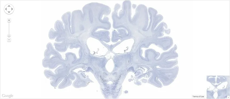
H.M.'s brain was imaged while he was alive using MRI and other techniques, but the new high-resolution model—created with data taken from photographs of the thousands of thin slices—has allowed the researchers to delve deeper into the brain's anatomy and make these sorts of observations on a finer scale.
They've discovered that some parts of the brain that were believed to have been left intact after the surgery were actually removed. The left orbitofrontal cortex, for instance, contained a small lesion, likely caused during the surgery. Additionally, they found that some portions of the left and right hippocampi were actually undamaged, a finding that could cause researchers to re-examine previous beliefs about the role of the hippocampus in different sorts of memory.
The UC San Diego team also plans to publish a free online "atlas" of the brain, made up of the high-resolution images taken of its slices, viewable on a zoomable Google Maps-like platform (one photo has already been published). Given that the original dissection of the brain was broadcast live on the web and attracted an estimated 400,000 viewers, it seems likely that in death, as well as life, H.M.'s extraordinary condition will captivate many.
/https://tf-cmsv2-smithsonianmag-media.s3.amazonaws.com/accounts/headshot/joseph-stromberg-240.jpg)