Blood Clots, Liver Cells and Bird Flu Are Surprisingly Beautiful Under a Microscope
The brightly-colored micrographs and scans in a new book, Science is Beautiful, answer big questions about the human body
/https://tf-cmsv2-smithsonianmag-media.s3.amazonaws.com/accounts/headshot/megan.png)
Most people know about ear drums—but what about ear stones? In the inner ear, tiny calcium carbonate crystals clump together to form stones called otoliths, which rest atop tiny hairs. When a person's head moves, so do these stones and the hairs attached to them. The hairs send impulses to the brain, and the brain in turn uses these signals to keep the body balanced.
Colin Salter's new book Science is Beautiful: The Human Body Under the Microscope features a scanning electron micrograph of the bumpy surface of one of these stones, its many crystals artificially colored pink, purple and blue. It's among myriad examples of vivid micrographs and MRI scans the science writer has curated to show cells, blood vessels, organs and diseases from a unique and sometimes startling perspective. The collection highlights "images of elements within the body whose existence you may never have pondered but whose functions are vital and fascinating," says Salter.
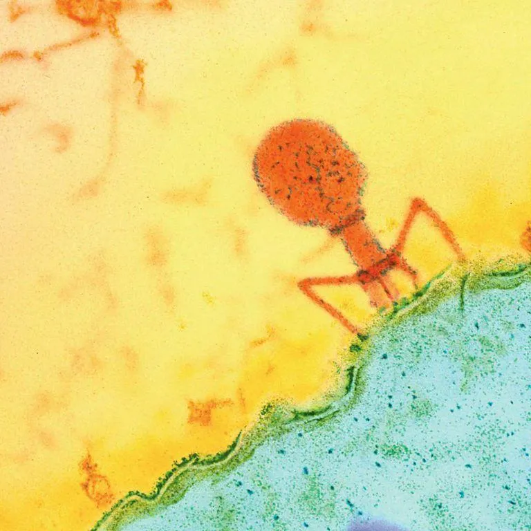
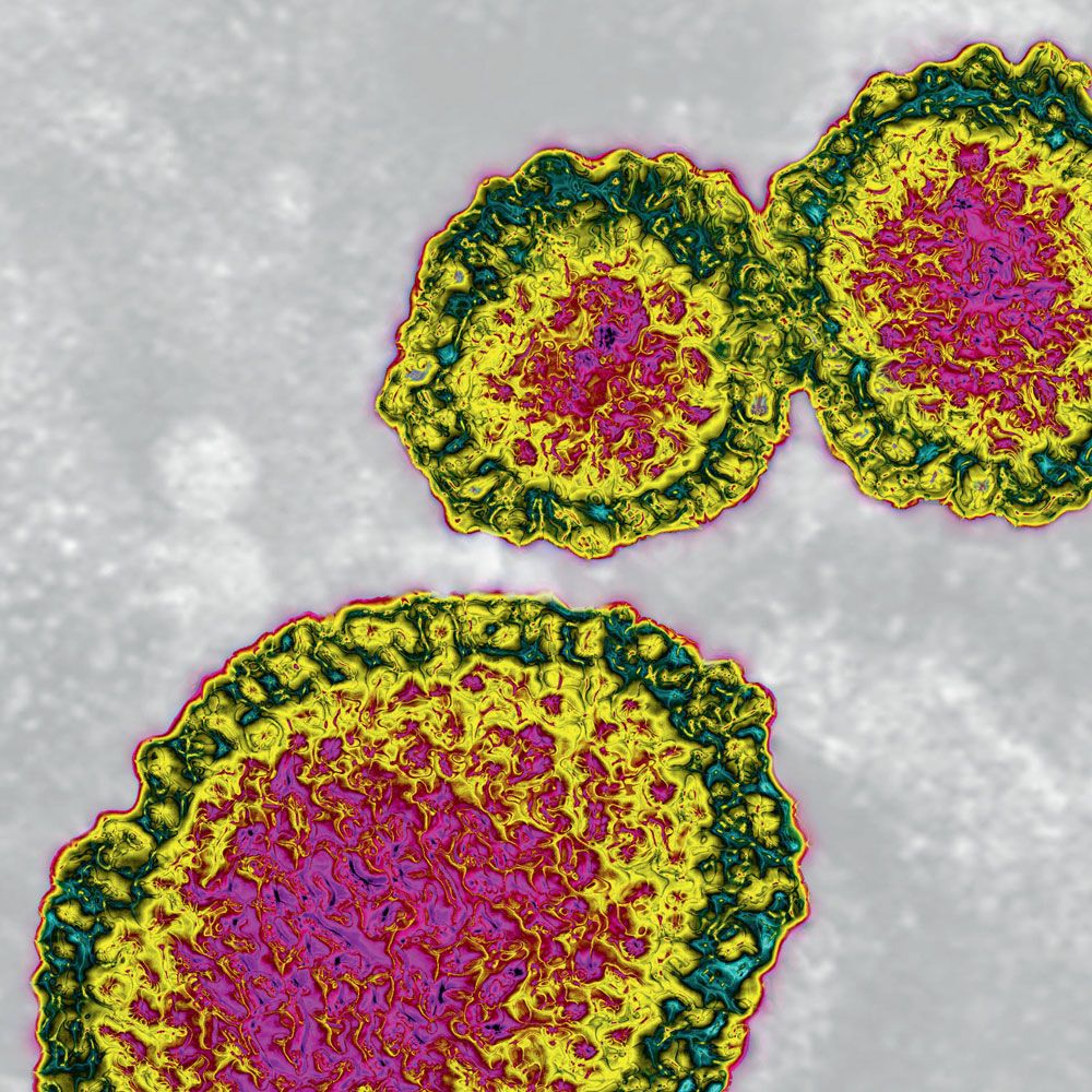
For most of the images, biologists stained the samples or added colors digitally to highlight various parts for their research and for other viewers. A dendritic cell looks like a pale pink peony in an ion-abrasion scanning electron micrograph. And while H1N1, or bird flu, has nasty effects on those who contract it, the virus itself becomes a spectacular mosaic in a filtered transmission electron micrograph. Here's what Salter had to say about the project:
What inspired you to write this book?
I have often written about the ingenuity of mankind’s scientific mind, our capacity for technical invention and innovation. But the most extraordinary machine of all is the one we inhabit—the human body.
With this book, I wanted to convey some of the wonder I feel about the complex systems that operate inside us all; repair and regenerate us; move both deliberately and instinctively, with and without conscious thought; learn how to do things better; and defend us from viruses, bacteria and just about anything we can throw at our digestive system.
What do you personally find compelling about micrographs of the human body?
Even without knowing what you’re looking at, they are beautiful. I’m fascinated by that: the idea that beauty doesn’t depend on meaning. The same shapes and colors can be beautiful whether they’re in an artery or in an art gallery.
But when you add meaning to beauty, when you add understanding to that instinctive visual pleasure, it greatly enhances your sense of wonder. Looking at these vivid organic shapes and intense colors and knowing that they show what’s going on inside of me, I am in awe.
Science Is Beautiful: The Human Body Under the Microscope
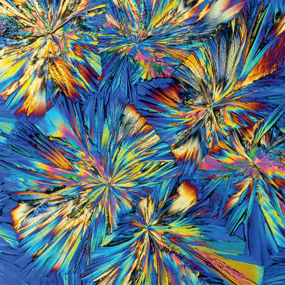
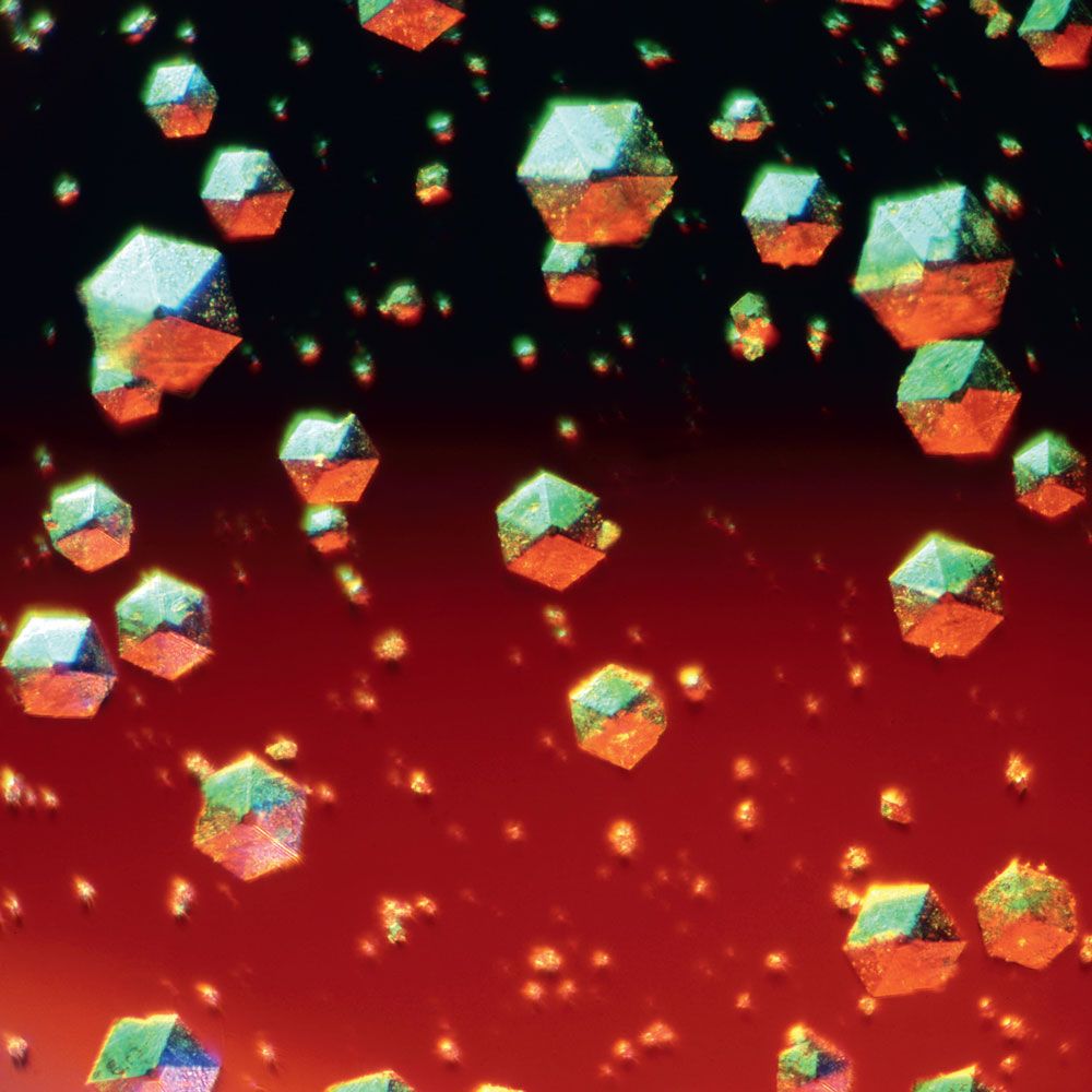
Take a fantastic journey through the human body, thanks to the magic of technology and the most spectacular microscopic images ever created. These pictures, as beautiful as any art, provide a window into the wonder of our brains, the work of a white blood cell, the power of hormones, the tiny hairs on our arms, the movement of human cancer cells, the jagged edges of caffeine crystals, and more.
What is your favorite image in the book, and why?
There’s a very pretty artificially colored image of hundreds of E. coli bacteria, which look like the sort of tasty candy you could scoop up and eat in handfuls! The picture of the cells that line the lungs is strikingly graceful. The structural skeletons of the cells appear as spun gossamer and make me think they are dancing some fantastic air ballet.
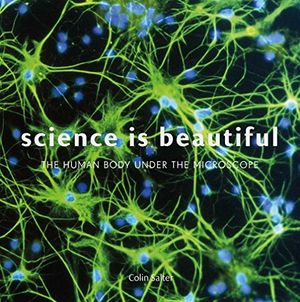
But I think my favorite is a rather stark black-and-white image of nerve cells in the cerebral cortex. They are responsible for conscious thought, memory and language, which is in itself a wonderful thought for a writer. But the picture looks like a forest of bare trees in a snowy landscape and speaks to my northern soul. I am Scottish, and we Scots are born and raised in wind and rain, in a land of short summers and dark winters. I can picture myself happily hiking through that forest.
What was the most interesting thing that you learned about the human body in the making of the book?
I had never heard of Henrietta Lacks, who suffered from cervical cancer and died in 1951. The cancer cells removed from her body proved to be particularly durable, and in a sustaining laboratory environment, they are still able to divide and multiply to this day. They are known as HeLa cells in Ms. Lacks’ honor. Although there are ethical questions about their continuing growth and use, it’s a kind of immortality, and this precious resource offers scientists a stable basis for decades of ongoing research into cancer treatments. There are two micrographs of HeLa cells in the book.
What do you hope readers take away from it?
Most of us don’t give much thought to what goes on inside us, which is probably just as well—not out of squeamishness, but because the complexity of it all is overwhelming. In Science Is Beautiful, I haven’t written a medical textbook, but I hope the book presents some strikingly beautiful images with enough simple explanation for readers to be able to say, “Wow! That’s amazing.” Because it is, it really is.
/https://tf-cmsv2-smithsonianmag-media.s3.amazonaws.com/accounts/headshot/megan.png)