“VirusCam” Can Watch Individual Viruses to (Someday) Keep You From Getting Sick
Viruses are tiny and hard to see, but a new microscope can track them individually to try to better prevent disease
/https://tf-cmsv2-smithsonianmag-media.s3.amazonaws.com/accounts/headshot/matchar.png)
/https://tf-cmsv2-smithsonianmag-media.s3.amazonaws.com/filer/b6/dc/b6dc2a2e-3bba-4991-94ba-b507204fc904/istock-610750282.jpg)
Viruses are small. Really small. Some are 1,000 times tinier than the diameter of a human hair. Once they’ve attacked and attached to a cell, they tend to move slowly, which makes it possible to see them under an electron microscope. But before that, when they’re all on their own, they’re just little chunks of genetic material in a protein coat, wriggling in unpredictable patterns, making them almost impossible to track. This has long been a problem for virologists, who want to track viruses to better understand their behavior.
Now, researchers at Duke University have developed a way to do just that – watch unattached viruses moving around in real time. This “virus cam” could give insight into how viruses break into cells, potentially giving rise to new ways of preventing infections.
“What we’re trying to do is figure out how viruses behave before they interact with cells or tissue, so we can potentially find new ways to interrupt the infection process,” says Kevin Welsher, the chemist who lead the research. The findings were recently published in the journal Optics Letters.
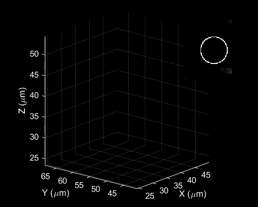
A video by the virus cam represents the path of a lentivirus, part of a group of viruses that cause deadly diseases in humans, as it moves through a salt water solution, traveling in an area barely wider than a human hair. The color changes in the video represent the passage of time – blue at the beginning, moving to red at the end.
The behavior of unattached viruses is “kind of an unexplored territory,” Welsher says. He likens trying to watch an unattached virus in action to tracking a high-speed car chase with a satellite.
“Your virus is a little car, and you’re taking satellite images and refreshing them as fast as you can,” he says. “But you don’t know what happens in between, because you’re limited by your refresh rate.”
The virus cam is more like a helicopter, he says. It can actually lock in on the virus’s position and watch it continuously. The camera was built by Duke postdoctoral researcher Shangguo Hou, who rigged a microscope to use a laser to track the virus so it can be held in view by the microscope’s platform, which is designed to respond very quickly to the optical feedback from the laser.
The virus cam is exciting because it can lock in on the virus’s position, Welsher says, but right now that’s all it does. Furthering the car chase analogy, he likens the virus cam to a helicopter that follows a car but can’t see any of its surroundings – the road, buildings, other cars. Their next step is to move beyond simply tracking the virus’s position to trying to understand its environment. Welsher and his team would like to integrate the virus cam with 3D imaging of cell surfaces, to see how viruses interact with cells before attempting to penetrate them.
This isn’t the first time researchers have caught individual particles moving in real time. Three years ago, while at Princeton, Welsher himself developed a method of tracking a virus-like fluorescent bead made of plastic nanoparticles moving into a cell membrane.
Viruses are more difficult to track than the beads because, unlike the bead, viruses don’t give off any light by themselves. Tagging viruses with fluorescent particles makes the viruses easier to see, but those particles are so much larger than the viruses themselves they likely interfere with the way the viruses move and infect cells, according to Welsher. The new microscope, because of the optical feedback provided by the laser, can detect the very faint light given off by tiny fluorescent proteins, which are much smaller than the virus. So Welsher and his team inserted a yellow fluorescent protein into the virus’s genome to allow it to be tracked without changing the way it moves.
Scientists have also come up with other ways to track very small things. One team used algorithms to track viruses, training their microscopes on where the algorithms predicted the viruses would be. In recent years, British researchers also developed an incredibly sensitive optical microscope that can see structures as small as 50 nanometers across, as small as many viruses. This allows them to see viruses doing their work inside living cells, whereas electron microscopes can only be used for dead, specially prepared cells.
Once chemists understand more about how viruses interact with cells, virologists and molecular biologists could get involved to see how their behavior might be manipulated, perhaps stopping them before they infect a healthy cell.
“The ideal scenario is we uncover some insight that’s actionable,” Welsher says.
/https://tf-cmsv2-smithsonianmag-media.s3.amazonaws.com/accounts/headshot/matchar.png)