How the Smithsonian’s Coelacanth Lost Its Brain and Got It Back Again
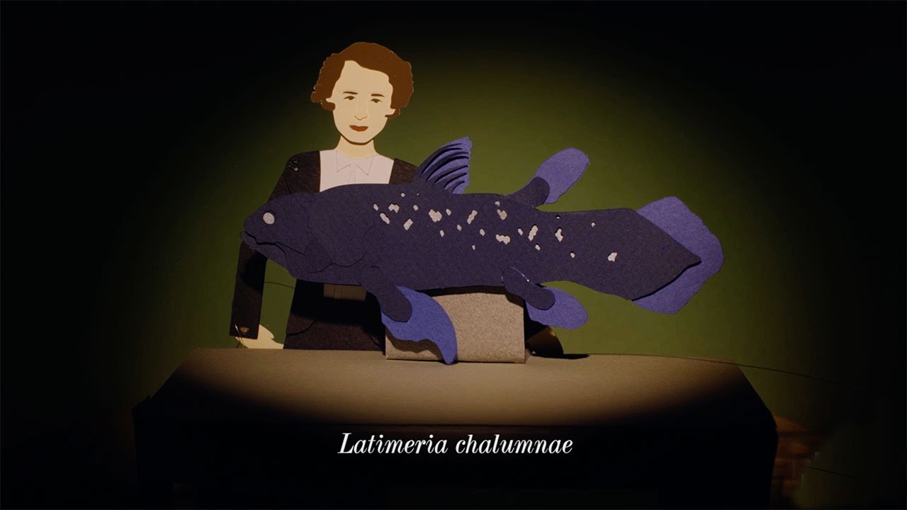
This year marks the 80th anniversary of the discovery of a fish believed to have gone the way of the dinosaurs 70 million years ago
/https://tf-cmsv2-smithsonianmag-media.s3.amazonaws.com/accounts/headshot/Parenti_Coconut_Island_2013.JPG)
/https://tf-cmsv2-smithsonianmag-media.s3.amazonaws.com/filer/5a/e1/5ae1369b-1943-4981-b8bb-a6b226761f3c/usnm_205871_latimeria_chalumnae_smith_photograph_right_side-wr.jpg)
Until 1938, finding a living coelacanth (pronounced “SEE-luh-kanth”) was considered as unlikely as seeing a velociraptor in your garden. The discovery of the long thought-to-be extinct fish off the Indian Ocean coast of South Africa 80 years ago this coming December was nothing short of astonishing. Textbooks of the time said this lineage of lobe-finned fishes, well known from fossils beginning in the Devonian period, about 360 million years ago, died off with the dinosaurs some 70 million years ago.
The story of the coelacanth’s discovery eight decades ago by a young woman, Marjorie Courtenay-Latimer, is told and retold, including recently in this delightful video by HHMI BioInteractive that recounts details of this remarkable find, and with the timeless appeal of puppetry and animation.
Living coelacanths were unknown to the scientific world for so long in part because of their limited distribution—in deep-water habitats in the western Indian Ocean and, as of 1997, deep off the coast of Sulawesi, Indonesia.
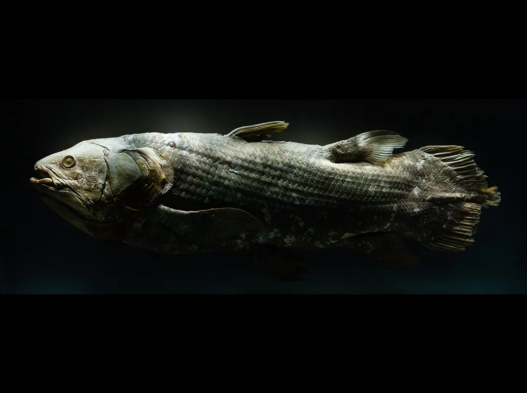
Once discovered, scientific collection proceeded at a modest pace and the number of specimens worldwide now totals about 300. In the Smithsonian’s National Collection of Fishes, at the National Museum of Natural History, resides one adult specimen of the coelacanth, Latimeria chalumnae, taken near the Comoros Islands off east Africa in the mid-1960s.
In mid-1968, the late Dr. H. Norman Schnitzlein (1927-2004), then a professor of anatomy at the University of Alabama Medical Center, purchased the coelacanth specimen for his research funded largely by the National Institutes of Health on the comparative anatomy of fish brains. By studying the form of fish brains, Schnitzlein and colleagues could better understand the brains of humans and other mammals. He described his philosophy for an NIH newsletter: “Our approach –the approach of comparative anatomy—is really a study of nature’s experiments on life and nature’s variations on the many forms which life takes.”
At the time of capture, the coelacanth specimen weighed about 160 pounds and measured a little less than five and a half feet long—average for adult coelacanths, which can reach 6 feet long, weigh as much as 200 pounds and live up to about 60 years.
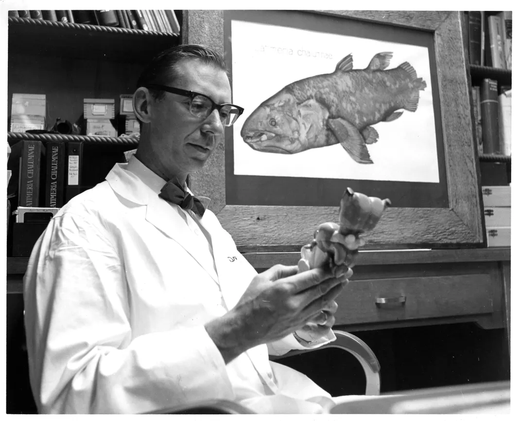
Collectors injected the specimen with formalin (a solution of formaldehyde) for preservation, and shipped it from east Africa to the U.S. Upon arrival at Schnitzlein’s lab, it had a formalin bath with some extra formalin poured directly onto the brain. Schnitzlein removed the brain for his own studies and donated the remainder of the specimen to the Smithsonian Institution in late 1968. A photograph of the museum’s coelacanth and some black and white line drawings of its brain grace a chapter written by Schnitzlein for a book on the anatomy of the vertebrate brain.
The coelacanth brain is small; it weighs about three grams. Sometime during its journey, someone gutted the Smithsonian’s specimen, possibly because it was starting to rot. Its sex is not known. The brainless, gutted specimen rests in a large tank filled with ethyl alcohol at the Smithsonian’s Museum Support Center in Suitland, Maryland. (Another on view in the museum’s Ocean Hall is a specimen on loan from South Africa.) Despite its history of gross dissection, the specimen is in good condition and is still a “must-see” for visitors to the collection.
When I look at it I see a survivor of a long-lived line of remarkable fishes. The living coelacanth is just like its 300-million-year-old fossil relatives with lobed-fins and a distinctive elongate middle portion of the caudal or tail fin. In life, coelacanths are a beautiful porcelain blue with distinctive cream-colored blotches. I dream of swimming with the coelacanths, but most of us will never see a live one as they dwell in the deep sea and are rarely taken at a depth less than 300 feet. This is good for the coelacanths as all are considered endangered.
Some years ago, the story of the Smithsonian’s coelacanth took an interesting twist. To study the anatomy of the brain, Schnitzlein’s lab employed a centuries-old technique: tissue histology. A histology technician would place a small piece of brain tissue in a bath of paraffin, a wax that makes it hold its natural shape, and cuts it to form a block. Then, using a special instrument, a microtome, the technician would slice the paraffin block into ultrathin sections, about 0.0002 inches thick. Finally, the technician would mount a thin glass coverslip over the slides to protect the sections. In this way, the magnifying and illuminating power of a microscope could then make individual tissues visible.
When Schnitzlein moved to the University of South Florida College of Medicine in 1973, he took his brain slide collection with him. When he retired in 1994, the USF medical school did not want to maintain his collections of fish brain slides. Schnitzlein gave the collection to Harry Grier, a research associate at the Natural History museum and a fish reproductive biologist at the Florida Fish and Wildlife Research lab in St. Petersburg, Florida. Grier put the slides in a cabinet for safe keeping.
But as the building they were in was scheduled to be demolished, someone else moved the boxes outside, destined for the dumpster. The Smithsonian had the coelacanth body, but not its brain and that was about to be trashed. Grier knew the museum would want this valuable collection and rescued it. In 1998, he packed and shipped the Schnitzlein collection—125 boxes of histological slides of fish brains, covering all major fish groups—to the Smithsonian.
The gem of the fish brain collection is the five boxes of histological slides of the brain of the museum’s coelacanth, numbered 1 to 3 and 5 to 6. Box number 4 is missing and presumed lost.
Grier also recovered and delivered to the Smithsonian a goldmine of letters, photos and other documents related to the original purchase of our coelacanth specimen. Today, the brain slide collection is undergoing an upgrade, overseen by museum specialist Jeff Clayton, to replace all the wooden boxes with new plastic ones.
The brain slides had faded after decades in storage and were difficult to use or interpret. Fortunately, a faded histological slide can be revived. As part of the upgrade, the museum’s histologist Helen Wimer simply reverses the original procedure. The coverslip is soaked off. The “baked-on” slices of paraffin-infused tissue on the slides can take up new stain, and then get a new glass coverslip. The refurbished slides are as good as new and now ready for the next generation of scholars of brain anatomy. Both the body and the brain can remain “in perpetuity” as scientific specimens in the National Collection of Fishes.
Editor's Note 9/14/2018: A letter arrived from the son of Dr. Norman Schnitzlein and we reprint it here for our readers.
Dear Ms. Parenti,
Thank you for your article about the coelacanth. Dr. H. Norman Schnitzlein was my father and, with your many shared research interests, I believe, had you known him, you and he would have been great friends. I forwarded your article to my siblings as it brought back many fond memories. We especially enjoyed the photo of our father in the article which none of us recalled seeing before.
Please allow me to share some some background about my dad's acquisition of the coelacanth. . . It was the holy grail of his research career of fishes. When he took me fishing as a boy, we'd always save the heads of whatever we caught in jars of formaldehyde for later removal of their brains. During his time at the University of Alabama in Birmingham he obtained all sorts of fish specimens. He was especially interested in the many varieties of lung fish. Also around the time he had the coelacanth, there were two electric eels swimming in a tank in his office that were destined for a similar fate.
After months of letter writing to Madagascar, the coelacanth was finally on its way to Birmingham, but somewhere en route it got lost. Weeks past when it was to have arrived, I recall him on the phone anxiously trying to track down its whereabouts. T
he coelacanth was finally found on a loading dock at the Atlanta Airport. Apparently, the coffin-sized box it was shipped in smelled terrible and likely contained something dead so no one wanted to touch it.
When the coelacanth finally arrived, everyone wanted to see it. I recall my dad opening this large metal box to expose this beast covered in formaldehyde-soaked cheesecloth. One of his less-bright students asked where he got the enormous fish. I'll never forget my dad, in dead-pan humor, saying he caught the coelacanth with a "pink-eyed hell-bender" lure up on Lake Purdy just outside Birmingham. A man of many talents, my dad made the picture frame from salvaged wood that held the image of the coelacanth in the background of your article's photo.
Thank you for indulging me with this trip down memory lane with my dad's coelacanth. And thank you again for mentioning him in your article. We still miss him.
Best Regards,
Paul Norman Schnitzlein
/https://tf-cmsv2-smithsonianmag-media.s3.amazonaws.com/accounts/headshot/Parenti_Coconut_Island_2013.JPG)