Inside a Brain Bank, Where Humans’ Most Precious Organ Is Dissected and Studied
Unlike organ transplants, brains are used primarily to support research of some of the most widespread and debilitating diseases in the world
/https://tf-cmsv2-smithsonianmag-media.s3.amazonaws.com/filer/01/89/0189d846-632c-475b-b06a-8d0bf3cea8c4/dsc_7984.jpg)
Around three or four in the morning, Tina Zheng leaves home to meet a brain. “I’ll try to nap a little bit in the Uber ride, and then I’ll review all the brain regions in the car ride too,” she says. “We’re never sitting down doing a boring office day job. It’s just the next second, there’s a brain coming, and we have to be up and ready for it.”
Zheng works as a tissue coordinator at the Harvard Brain Tissue Resource Center (HBTRC) at McLean Hospital, one of the oldest brain donation banks in the country. Brain matter has a limited shelf life, so dissectionists like Zheng are on-call around the clock to partition and preserve a freshly donated brain as soon as it arrives to the lab, whatever time that may be.
Unlike other organ donations, which are generally used for transplants, brains are primarily harvested to support the research of neurological diseases and disorders. Every year, the HBTRC sends thousands of brain tissue samples to labs all over the world to investigate the causes and possible treatments of ailments such as addiction, Alzheimer’s and Parkinson’s.
Research supported by brain banks is increasingly critical with today’s rising rates of neurological illnesses. Between 1990 and 2015, the number of deaths from neurological disorders, including stroke, dementia and multiple sclerosis, increased by nearly 37 percent. In the United States alone, 5.8 million people currently live with Alzheimer’s, making it the nation’s sixth leading cause of death. By 2050, the affected population is projected to increase to almost 14 million.
Although treatments for these disorders remain elusive, postmortem brain tissue offers a key resource for unlocking possible solutions. In the past, neurological medications like lithium, used to treat ailments such as bipolar disorder, were discovered mostly by trial and error, with researchers or physicians prescribing different substances and observing the effects. Thanks to advances in genetics and biotechnology, scientists can now hunt for specific genes and molecular pathways that are related to a particular disease, and hopefully find treatments that are tailored to these targets. Brain tissue serves as the raw material for exploring genetic and environmental factors of neurological conditions, making it a critical substance for meeting the needs of brain research labs around the world.
“It’s that feeling of, I am contributing to something much more than myself that keeps me going,” Zheng says. “The human brain, there’s so much mystery involved in it right now, I want to be part of discovering what’s next and what’s left to uncover.”
**********
The brain is like a ticking time bomb whose clock starts at the moment of death. If a brain isn’t retrieved within 24 hours, its texture changes from a “firm Jello” to “pudding,” Zheng says, rendering it essentially useless for research. Given this short window of time, the brain donation team must work as efficiently as a fine-tuned assembly line.
The brain donation process starts when a donor is near death, or soon after they pass away. The family notifies a coordinator at the HBTRC, who secures a pathologist to remove the brain and a courier to retrieve it.
“Most of the time it’s a cold call,” says Kay Sullivan, program manager at the HBTRC. The coordinators screen potential donors for issues like massive strokes or infectious diseases, and direct the families to place the bodies in refrigeration within six hours. Most brain removals take place in a funeral home rather than a hospital, since hospitals are wary of taking in “random bodies,” Sullivan says.
Allison Rodgers, another tissue coordinator, recalls her first week on the job when the brain bank received eight cases in just five days over Christmas. “Sometimes you’re working a case, and you’re wrapping up … just getting back to bed, then you get another phone call,” she says. “Honestly, all you do is put on another pot of coffee and keep going.”
The HBTRC accepts donations from every state in the U.S., so sometimes a pathologist or courier needs to drive several hours to retrieve a brain. Fortunately, the center has a network of over 500 pathologists across the country, making most brain donations possible.
Katherine Waters, the chief of pathology and laboratory medicine at VA Maine Healthcare, started harvesting brains for the HBTRC this year. “When I get a call saying where the death is, I get the kit, get my car, get to the funeral home, and harvest the brain,” Waters says.
Waters’ brain removal kit is like something Florence Nightingale might carry if she were also a mechanic and a superhero. It contains a fluid-impermeable jumpsuit, a catheter to extract cerebrospinal fluid from the spinal cord, a tool resembling a chisel to pull off the calvaria, or skullcap, and a bone saw, which is similar to an electric cast saw. “You could do it by hand,” Waters says of cutting open a skull, “but it would take a lot more time, and I don’t have the physical strength to do that.”
Waters starts by elevating the upper body and making an incision from the back of one ear across to the other, allowing her to pull away the scalp. Using the bone saw, she cuts a circle across the skull and twists the skullcap off with the chisel. With the skull open, Waters can collect fluid samples and then remove the brain, placing it in a bag with a slurry of ice, and then in another bag and a Styrofoam cooler. Afterwards, she reassembles the skullcap and scalp as if nothing ever happened. The whole process takes less than an hour.
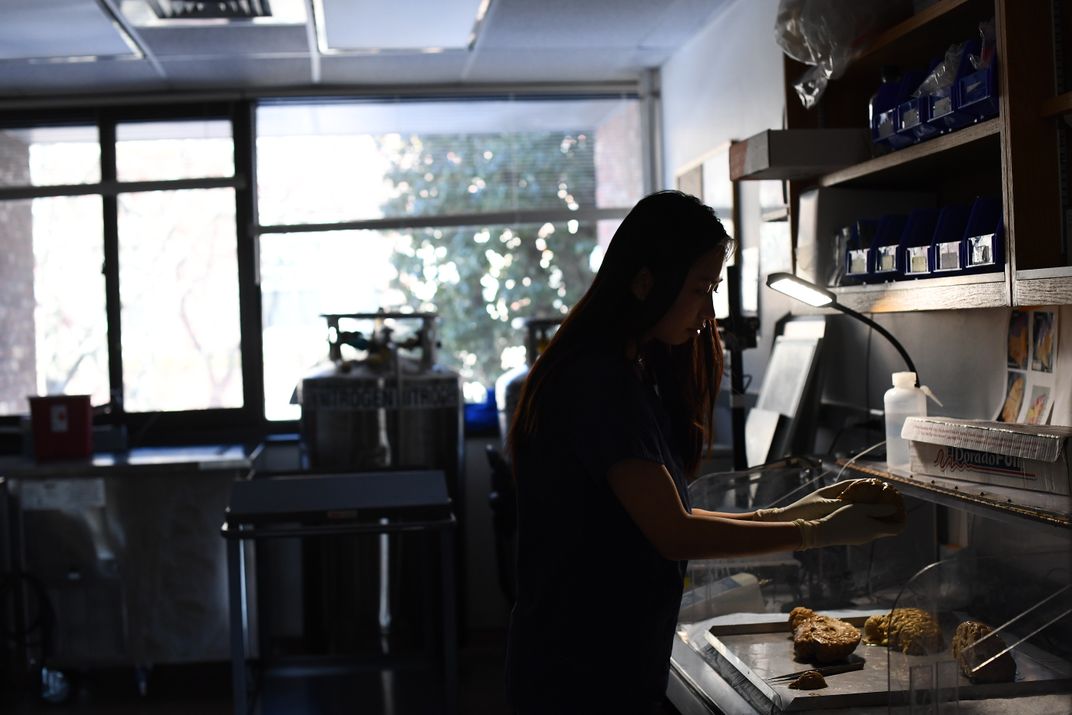
The brain makes its way from the funeral home to the HBTRC via a courier, sometimes on a commercial flight. Team members like Zheng and Rodgers receive notice of the brain’s arrival and assemble at the brain bank to perform the dissection. Like Waters, they follow a strict routine.
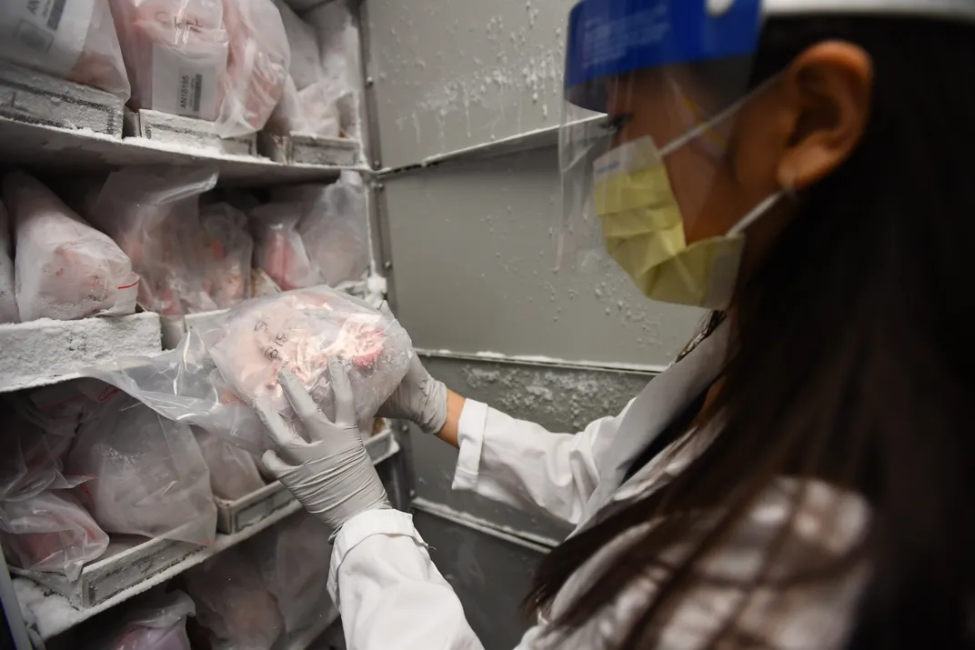
After weighing and photographing the brain to note any abnormalities, the dissectionists cut it in half. One side is further dissected and frozen at minus 80 degrees Celsius, while the other is fixed in formalin. Throughout each of these steps, dissectionists look for signs of disease progression, like the shrunken frontal lobe that can accompany dementia or the deteriorated striatum of Huntington’s disease.
“Throughout the whole process, we can see how much every part of the brain changed based on what the person was experiencing,” Zheng says. “You can kind of imagine what the person’s life was like and how the disease really affected him or her.”
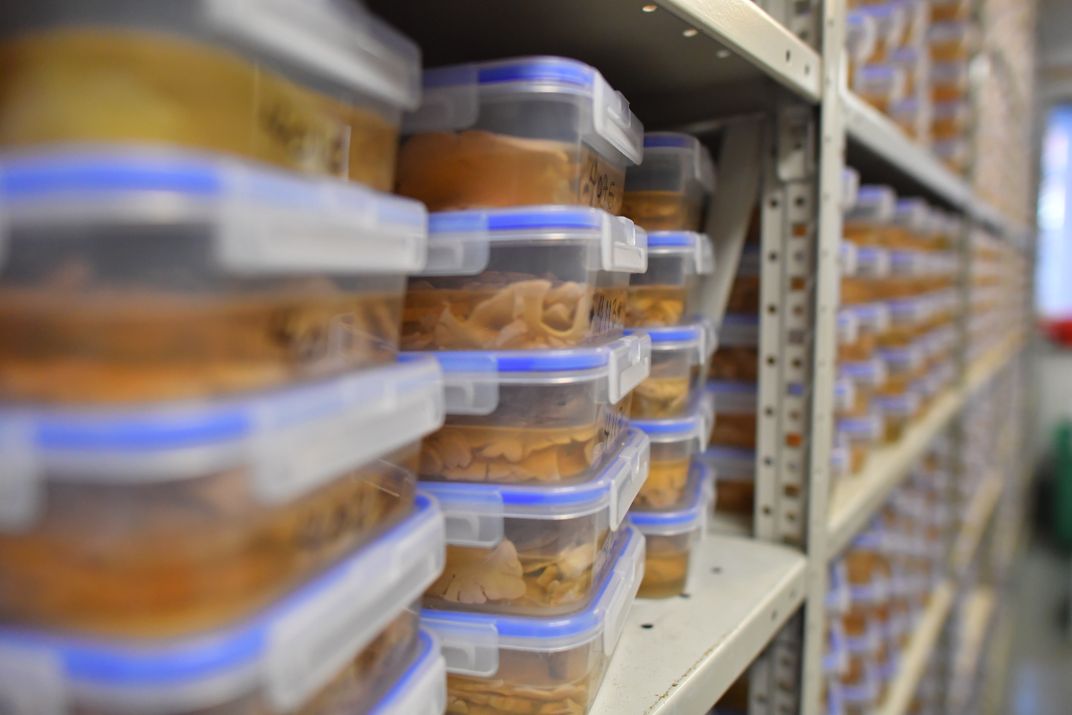
After about three hours, the dissection is complete. The formalin sample joins over 6,000 other half-brains in the “fixed room,” and sections are made into slides and stains that histologists examine under a microscope to look for abnormalities in the tissue, like plaques characteristic of Alzheimer’s. Scientists from around the world can later request samples that match the specific pathology of their research.
“I don’t think that feeling you get when you first pick up a brain ever goes away,” Zheng says. “I feel very privileged to be in this position and to be able to talk to their families and hear their stories, and to be trusted with their loved ones’ tissue. … Seeing the tissue in my hand and cutting it and thinking about the person’s life—I think it’s really empowering.”
**********
Postmortem samples from brain banks like the HBTRC have already started to support breakthroughs in neurological research. For instance, a recent study using tissue from the cortex of donated brains pinpointed specific neurological pathways that are affected by autism spectrum disorder. Other work used donated tissue to look at the genes associated with major depressive disorder. The scientists found that the expression of genes varied based on factors like gender, providing key information that could someday be used to develop more efficient and personalized antidepressant treatments.
“You need all the tools you can get to understand these disorders,” says Sabina Berretta, director of the HBTRC. In addition to studying postmortem tissue, imaging methods like MRIs offer alternative tools for investigating neurological conditions.
Berretta explains that while imaging has the advantage of allowing scientists to study living subjects, it has poor resolution (“maybe a square centimeter”), and “you only get a slice of a person’s life at a certain time of their disorder.” By contrast, postmortem tissue provides resolution at the molecular level and could reveal lifelong patterns in a subject’s history. The two techniques are complementary, allowing broad analysis of living brains and then a more meticulous investigation of donated tissue.
“I like to think of it as if you were exploring a completely different part of the world,” Berretta says. “You would first want to fly at high altitude—get a bird’s eye view ... but if you want to know about the plants and animals, what language they speak there, what houses they live in, you need to go to the ground.”
**********
Even with teams working around the clock, brain banks are sorely lacking one critical component: healthy brains. While people suffering from neurological disorders are more likely to register as donors to contribute to a future cure, healthy people usually don’t think to donate their brains. This absence places an enormous limitation on research, since scientists need control samples to compare against diseased tissue.
“I think a lot of people are scared of brain donation because it involves death, and a lot of people, especially young people, don’t plan their funeral,” Sullivan says. “There is a stigma … so people are scared of the topic.”
Sullivan cites some common misconceptions, such as the idea that brain donation prevents a funeral viewing (it doesn’t, as cuts are only visible on the back of the head). Many also assume that the brain is included in the standard organ donation you sign up for on your driver’s license, which prioritizes transplant and only retrieves the brain after it’s started to degrade.
“I think in the future, we’re hoping there will be a database where you can select which body parts will go to research, and then if there’s a car accident or something, you’ve already given pre-mortem consent for [brain donation],” Sullivan says.
Efforts to increase the number of brain donations are already under way. Tish Hevel founded the Brain Donor Project in 2016 to help spread the word about the NIH’s NeuroBioBank, a national network of six brain banks including the HBTRC that was established in 2013. Motivated by a nightmarish experience trying to facilitate her father’s brain donation while he was suffering from Lewy Bodies dementia, Hevel started the nonprofit to ease the process of connecting donors with brain banks.
"There are more than 50 million Americans [with neurological disorders],” Hevel says. “That's [almost] one in five of us ... and we're not getting answers fast enough. There simply is no substitute for human brain tissue. Many neurological researchers say that is the most precious substance known to man."
In its first two years of operation, the NeuroBioBank supplied more than 10,000 tissue specimens to support almost $70 million in research funding that involved postmortem brain research. Since October 2016, the Brain Donor Project helped reach more than 6,300 new donors from all fifty states. But Hevel emphasizes that there is still a long way to go.
“When we were first setting up, [experts asked], ‘What if we’re too successful? What if we have too many brains?’” Hevel says. “It’s just not going to happen in our lifetime. It’s just such a critical issue, [and] we’re just not making progress in key areas. … We got to get to it so that people don’t have their whole families’ lives ruined.”
In light of this escalating need for neurological research, Hevel’s efforts offer hope: We might finally save the human brain from its most pervasive threats, if we choose to put our minds to good use.