How Studying Bioluminescent Creatures Is Transforming Medical Science
The natural light of insects and sea creatures can help doctors illuminate H.I.V. and even kill cancer cells
/https://tf-cmsv2-smithsonianmag-media.s3.amazonaws.com/filer/45/52/455201dc-16d2-4604-9e6c-2ebf1a3abc17/gettyimages-590266104.jpg)
When Cassius Stevani saw blue light emanating from the fallen branches in Brazil’s Atlantic Forest, he knew it couldn’t be coming from the bioluminescent mushrooms he was collecting. The University of São Paulo biochemist was working on a study of bioluminescence and photochemistry—the chemistry of light—when he and a team of researchers discovered Neoceroplatus betaryiensis, a new species of fungus gnat and the first insect in South America to emit blue light.
“It’s an important find for the areas of entomology, ecology, bioluminescence and evolution,” Stevani says.
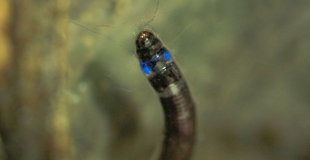
The larvae of the tiny flying creature, stuck to the branches and trunks of forest trees thanks to their own secreted silk, glowed from their tops and their bottoms, with one light in their last abdominal segment and another two on either side of their first thoracic segment, just under their heads.
The reason the gnats glow is still a mystery, but researchers hope its light continue to help them save lives.
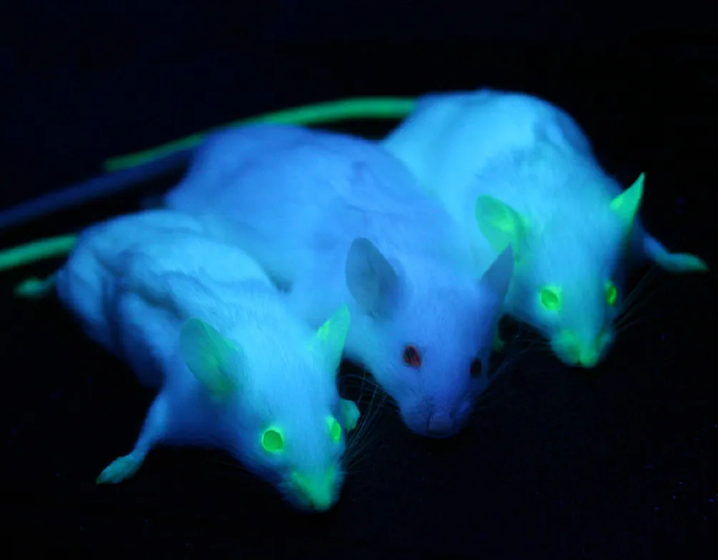
Bioluminescence comes in a range of greens, reds and blues, and it’s caused by a protein called luciferin, often found in marine animals, mushrooms, insects, algae and specific types of bacteria. In 2008, three scientists were awarded the Nobel Prize in Chemistry for their work with bioluminescence. They discovered, developed and genetically modified green fluorescent protein (GFP), making it possible for animals that don’t naturally glow to produce their own light. The work opened the door to a number of scientific applications, including the advancement of groundbreaking medical research.
GFP is now used as an important tagging tool in bioscience and can be attached to otherwise invisible proteins, allowing researchers to better understand cell damage in Alzheimer’s and other neurological diseases, improve the detection of blood clots, track the spread of H.I.V. and its transmission path, and even fight cancer.
“We in science should take more examples from nature,” says Theodossis Theodossiou, a senior researcher at the Institute for Cancer Research at Oslo University Hospital who uses bioluminescence to develop possible new treatments. “Nature creates systems that our technology is yet unable to create. When we see species that create light and do it chemically, it’s amazing. It’s a source of inspiration. The only thing we can do up to now is borrow these systems from nature, translate them into our systems, our research and our needs.”
Theodossiou had long been working on photodynamic therapy (PDT), a cancer treatment that uses bursts of laser light to attack tumors close to the skin’s surface. But PDT can’t be used to treat cancer hidden deeper in the body. So Theodossiou used the same molecules that create firefly light—a yellow-green glow—to develop bioluminescence-activated destruction of cancer (BLADe), a method that allows the light source to come from inside the cancer cells as opposed to an external laser.
After the cancer cell is treated with a photosensitizer—a molecule that causes a chemical change in other molecules after being injected into the bloodstream—the light triggers the cancer’s destruction. The BLADe technique, blasting cancer cells with light from within, essentially causes them to self-destruct, no matter how deep-lying the cancer or how far it has spread.
Theodossiou and his team have now identified photosensitizers that can be tailored to their research, allowing them to use not just to GFP to attack cancer cells, but also blue light—like that recently found by Stevani and his team in the fungus gnat larvae on the forest floor of Brazil.
Blue light was not previously used in PDT clinical trials because it wasn’t able to penetrate deeply enough into tissue. Now that bioluminescence can put that light inside cancer cells, a powerful photosensitizer only activated by blue light could be used to create a new cancer-destroying tool, and Theodossiou and his team are already working on such a tool.
“When we create the light from the inside, it doesn’t have to transfer any tissue; that’s the beauty of it,” he says. “We don’t care if the light is blue or yellow-green—as in the case of BLADe up to now—or red. We care that the photosensitizer is the most efficient one.”
For Thomas J. Hope, who pioneered the use of cell biology approaches to study H.I.V. at his laboratory at Northwestern University, that same photosensitizer efficiency is key to possible new treatments. His research into the transmission path of H.I.V. and how the virus interacts with other cells in the body led him to use bioluminescence from both fireflies and shrimp to tag and track S.I.V., a similar virus transmitted in macaques.
By tagging virus cells with bioluminescent proteins, Hope has made it possible to quickly find miniscule pieces of tissue, sometimes measuring just 1mm2, where S.I.V. or H.I.V. is passing and attacking other cells. These interactions can then be studied in greater detail than ever before.
“It’s the needle in the haystack problem,” he says. “If you need to find a needle in a haystack, how do you do that? If you can make it glow with a luciferase, then it’s much easier.”
Before bioluminescence helped H.I.V. researchers like Hope track the virus, similar work was done with radioactive materials, but the technique was much more expensive and significantly less safe. Luciferases—the enzymes that cause bioluminescence—are more sensitive and more amenable to use in a lab, requiring fewer precautions than working with radioactivity. Now, Hope and his team are also able to conduct tests on live animals, something that wasn’t possible with radioactive materials.
“It’s really given us a whole new tool, and I’m excited to see what the characteristics of this new luciferase might be so we can see where it might fill gaps in what we currently have,” he says of the discovery of the blue light-emitting fungus gnat. “Maybe it will have some very neat characteristics that could break open some new areas of research.”