Early Microscopes Revealed a New World of Tiny Living Things
A cloth merchant turned a device for checking his wares into an instrument fit for science
/https://tf-cmsv2-smithsonianmag-media.s3.amazonaws.com/filer/09/6c/096cd631-f91e-44ed-a0bd-5169548ca252/42-73760973.jpg)
Antoni van Leeuwenhoek had what some might consider an unusual hobby for a Dutch cloth merchant in the 17th century: making simple but exquisite microscopes.
His hometown of Delft in the Netherlands was experiencing a golden age of prosperity and cultural growth. The Dutch had recently won their independence from Spain, and the nation was rapidly becoming one of the wealthiest in the world, with a powerful navy and thriving international trade through the Dutch-East India Company. The newly wealthy became patrons of artists such as Rembrandt and Vermeer, and, freed from the constraints of Catholic Spain, scholars began to look at the natural world in a scientific way.
At the time, microscopes didn’t look anything like the ones now found in laboratories and classrooms, and they weren’t used much for science. Van Leeuwenhoek and other merchants used handheld microscopes to check their wares for flaws. But with time and money for leisure pursuits, van Leeuwenhoek began tinkering with these microscopes. And in the 1670s, he turned his devices to living things—and opened up a new world. He became the first person to observe the inner workings of the body on a microscopic level, seeing bacteria, sperm and even blood cells flowing through capillaries.
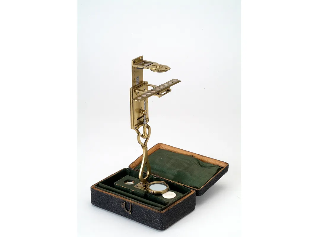
His microscopes, each smaller than the average thumb, “had a huge impact, and yet they look astonishingly simple,” says Marvin Bolt, curator of science and technology at the Corning Museum of Glass, where a rare van Leeuwenhoek microscope, on loan from the Museum Boerhaave in Leiden, Netherlands, is on display as part of an exhibition about the instruments.
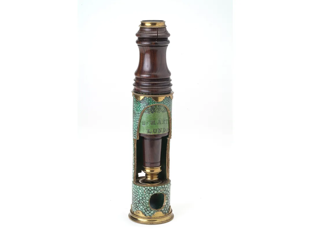
Lenses—curved pieces of glass that can focus light to create magnified images of objects—had been made in Europe and used for correcting vision since the 14th century. In the 16th century, Dutch lens makers began using high-quality Venetian glass to create lenses that produced clearer, sharper images than any before. Soon, someone used such a lens to create a simple microscope that could magnify objects. Then, a maker paired convex and concave lenses together, in an approach similar to how telescopes were made, creating the first compound microscope. By 1625, the term “microscope” had been born, appearing in a book by Italian scholars Francesco Stelluti and Federico Cesi, who had used the instrument to study honeybees.
Robert Hooke, an English scholar, also employed simple and compound microscopes to observe many aspects of the natural world, including fleas, plants and fungi. His Micrographia, the first popular science book, published in 1665, featured detailed engravings of flora and fauna as observed under microscopes with magnifications of roughly 20 times. Hooke also described how to make a simple microscope—inspiring van Leeuwenhoek and others.
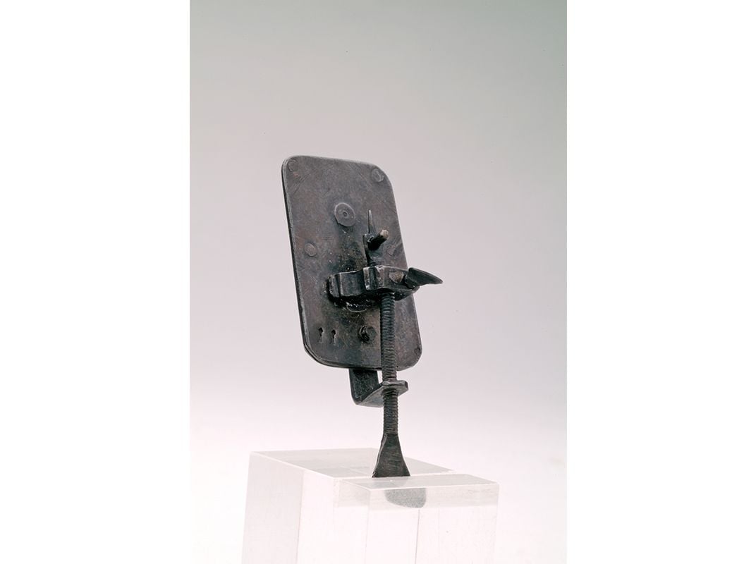
But van Leeuwenhoek took the burgeoning technology to new extremes, achieving higher magnifications than ever before: up to 300 times or so. He sandwiched a carefully crafted glass ball lens between the holes in two metal plates, which were riveted together. He then mounted the specimen on one side, on a needle that could be adjusted with the help of screws. The glass lenses were key, and van Leeuwenhoek used a few different techniques to craft his—and guarded his secrets closely.
In a compound microscope, like one found in a science lab today, a lens close to the object collects light to magnify the image, and then another lens in the eyepiece magnifies that image a second time. But the images in early compound microscopes were distorted. With a simple microscope, a single lens does all the work, and the specimen, lens and viewer’s eye are all very close together. In van Leeuwenhoek’s tiny contraption, the specimen was situated just millimeters away from the lens, producing a clear, sharp image for the viewer.
“As you increased the power, compound microscopes at the time were inferior to a good, simple lens instrument,” says Raymond Giordano, a historic microscope collector and dealer, and author of The Discoverer’s Lens: A Photographic History of the Simple Microscope, 1680-1880.
Van Leeuwenhoek examined samples he took from his own mouth and from water glasses and found them teeming with what he called “animalcules.” “When these animalcula or living Atoms did move, they put forth two little horns, continually moving themselves,” he wrote in the first scientific journal, Philosophical Transactions, after observing a sample of rainwater in 1675.
“Robert Hooke was looking at parts of animals that were already known,” says Bolt. “Then van Leeuwenhoek went deeper, to see, on a cellular level, things no one had ever seen before, such as muscle fibers, sperm and bacteria. He really blazed a trail.”
It was so difficult to bring a specimen into focus on his tiny instruments that van Leeuwenhoek usually made a microscope for each new specimen, some 500 devices in total, though only about a dozen originals are known to exist today. He gave away some and many were auctioned after his death, landing in various countries. Ultimately, though, it’s likely many were lost or melted down.
Van Leeuwenhoek’s findings were crucial to the scientific revolution and the development of the scientific method. But, like Galileo with the telescope, it would be almost 200 years before scientists such as Louis Pasteur would pick up where van Leeuwenhoek left off.
“Van Leeuwenhoek and his contemporaries were figuring out that they could discover things about the natural world not by reasoning, not by debating, but by actually observing and then confirming someone else’s observations,” says Bolt. “The priority of discovery was a new concept, as was replicability of scientific findings and objectivity.”
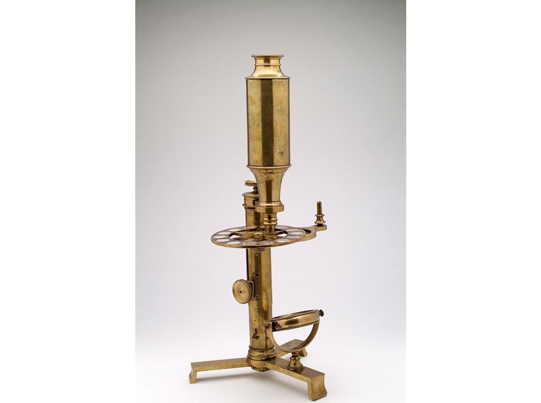
The simple microscope played an important role in science all the way up to the 19th century. Such microscopes “were long thought of as something only naturalists used,” Giordano recalls, noting that Charles Darwin used a simple microscope that he designed himself, but, in fact, all scientists of the time used them.
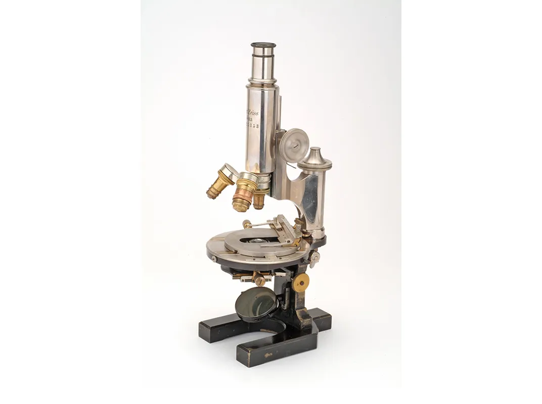
The 19th century brought major improvements to microscopes, including achromatic lenses, which allowed viewers to see color accurately for the first time. There were also new ways to illuminate specimens and control light, and the bases of compound microscopes became more stable. Finally, in the late 1800s, German chemists Otto Schott, Carl Zeiss and Ernst Abbe began scientifically engineering glass specifically for microscopes. By the late 1800s, microscopes were showing up in high schools.
Today, microscopes are more available than ever. The Internet is full of DIY tutorials for making a microscope by combining an iPhone camera with an inexpensive laser pointer lens. And last year, Stanford University introduced the Foldscope, a paper “print-and-fold” simple microscope that scholars believe could revolutionize global public health, science education and field-based citizen science. “It’s the logical conclusion to the history of microscopes, as instruments of knowledge,” says Bolt, “to get them from a few hands into many people’s hands.”
“Revealing the Invisible: The History of Glass and the Microscope” is on view through March 19, 2017, at the Corning Museum of Glass in New York.