This Painting Shows What It Might Look Like When Zika Infects a Cell
David S. Goodsell’s watercolor-and-ink artworks use the latest research to illustrate viruses, proteins and more
Zika virus exploded onto the global stage last year when health officials began to suspect it could cause birth defects in babies. Like the Ebola epidemic in 2014, fear burgeoned quickly. The destruction wrought by the disease is profoundly unsettling, in part because the particles of contagion are invisible.
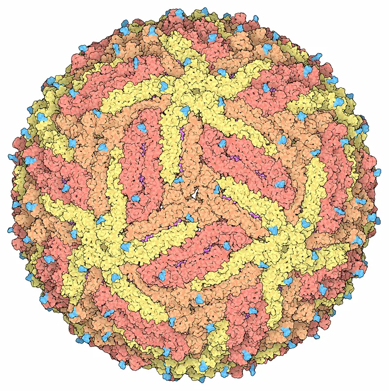
To make something visible is to get a better handle on it, to make it more manageable. In March of this year, Michael Rossmann of Purdue University in Indiana and his colleagues mapped what Meghan Rosen for Science News described as the "bumpy, golf ball-shaped structure" of Zika. With the structure deduced, scientists now have a starting point to learn how the virus works and whether it can be stopped. Researchers will look for points in the structure that might offer up a target for a drug.
In that vein, but with a more artistic twist, another scientist has painted an image of what it might look like when Zika infects a cell.
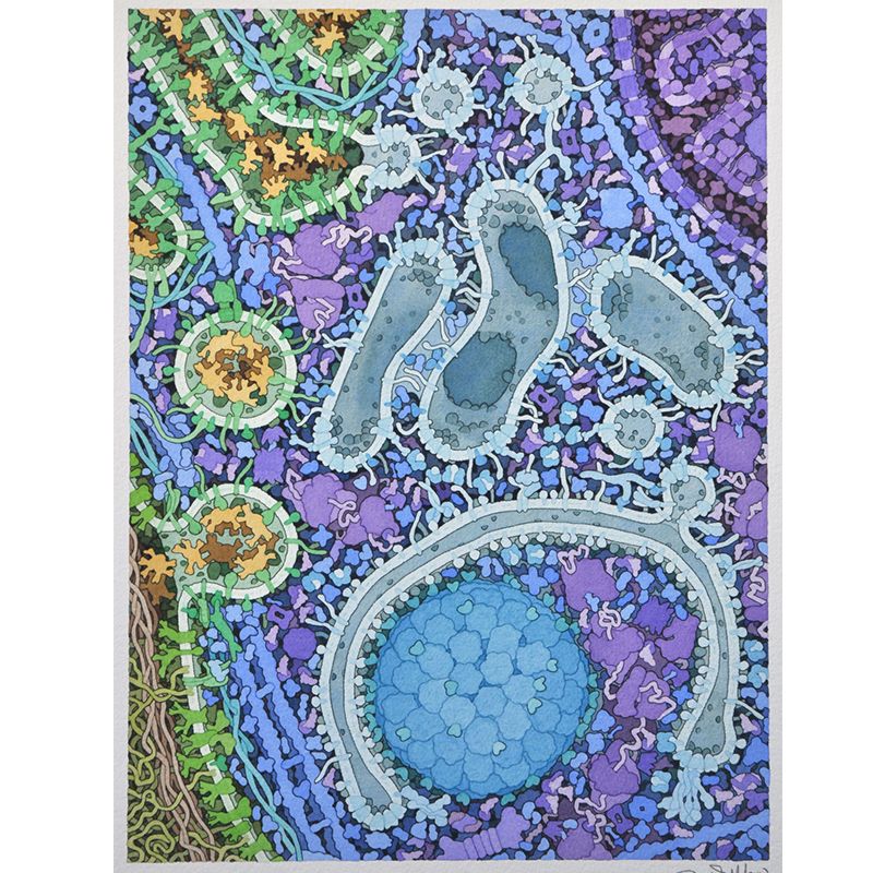
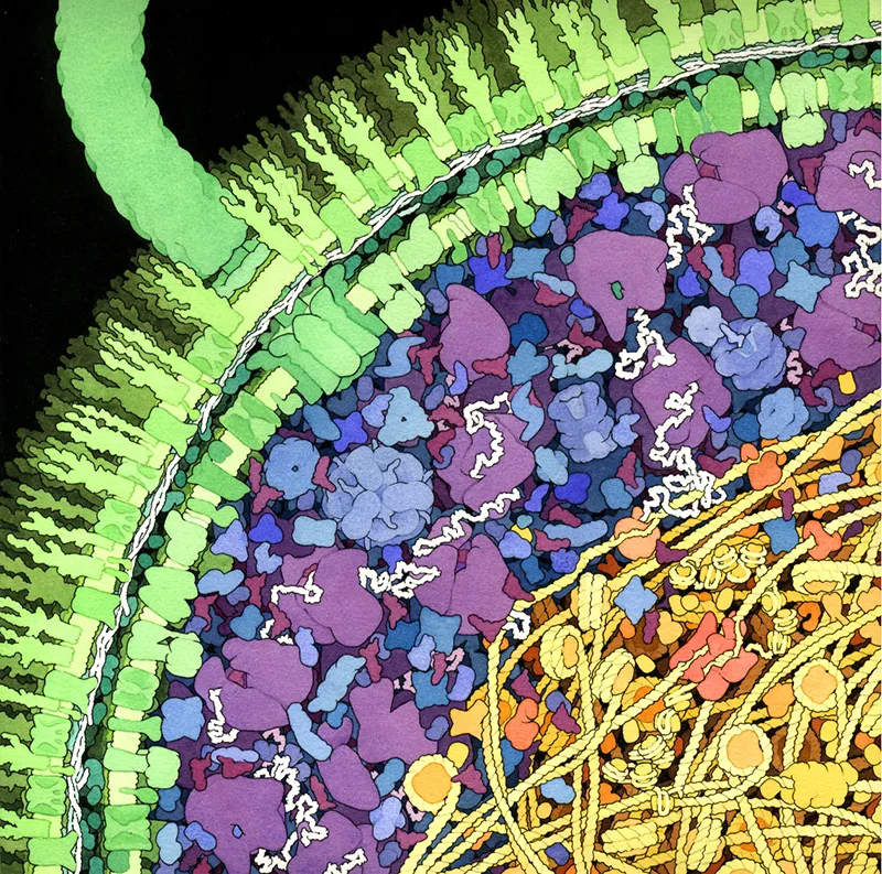
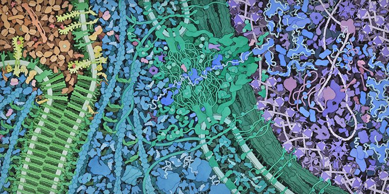
David S. Goodsell's watercolor depicts an area about 110 nanometers wide, reports Maggie Zackowitz for NPR. That's almost 1,000 times smaller than the width of a typical human hair. In the painting, a pink sphere representing the virus has been sliced in half to reveal tangles of the viral genetic material. Fleshy protuberances on the virus's surface grasp green towers embedded in a light green curve that seems to enclose a jumble of blue. The surface proteins of the virus are binding to receptors on the surface of a cell it will soon infect.
Deadly viruses never looked so beautiful as they do under Goodsell's brush. The molecular biologist with joint appointments at the Scripps Research Institute in La Jolla, California and Rutgers State University in New Jersey paints brightly colored and squishy-looking shapes resembling jellybeans, footballs and spaghetti that crowd and jumble together. As abstract images they are delightful, but Goodsell's work is also firmly footed in science.
The scientist-artist makes some educated guesses for his paintings. "Some of the objects and interactions are very well studied and others are not," he explains. "The science is still a growing field." But his expertise lets him wield the paintbrush with confidence.
Visualizing the microscopic biological world first intrigued Goodsell in graduate school, when he relied on techniques such as x-ray crystallography to deduce the folds, twists and contortions of proteins and nucleic acids.
Structure is key to giving molecules in cells their function, whether they are enzymes that cleave other molecules, RNA strands that instruct protein building or the fibers that support and shape tissues. Pockets in proteins offer up spots where other molecules can bind and catalyze or prevent reactions. When Rosalind Franklin succeeded in capturing the first picture of DNA, using x-ray crystallography, James Watson and Francis Crick were quickly able to deduce how unzipping the double helix could provide a template for replication of genetic material.
"If you are standing outside an automobile and the hood is closed so you can't see the engine, you have no idea how the machine works," says Stephen K. Burley, a researcher who studies proteomics at Rutgers University. Cells themselves are tiny, complex machines, and understanding how they work or what parts and processes go awry under the influence of disease, requires a look under the hood.
That's why Goodsell needed to understand how molecules were shaped as well as how they fit together inside the cell.
Computer graphics were just breaking into the research lab scene in the mid-1980s and giving scientists like Goodsell, now 55, an unprecedented look at the molecules they studied. But even the best programs struggled to show all the intricacies of a single molecule. "Objects the size of a protein were a real challenge," he says. Visualizing multiple proteins and their place relative to cellular structures was beyond the hardware and software capabilities at the time.
"I said to myself: What would it look like if we could blow up a portion of the cell and see the molecules?" Goodsell says. Without the high-powered computer graphic capabilities of today, he turned, quite literally, to the drawing board to piece together all the bits of knowledge about structure he could and create that image of the crowded interior of a cell. His goal was "to get back to looking at the big picture of science," he says.
The images he creates are meant to be scientific illustrations, to inspire researchers and the general public to think about the structures that underlay chemical reactions and cells' functions.
Typically, Goodsell spends a few hours digging through scientific literature to learn everything researchers know about the topic he wants to illustrate. Then, he draws up a big pencil sketch based on what he has learned. Carbon paper helps him transfer that sketch to watercolor paper. The molecules inside cells are often smaller than the wavelength of light, so a true view of a molecular landscape would be colorless, but Goodsell adds color and shading to help people interpret his paintings. The result is detailed views of molecular machinery at work.
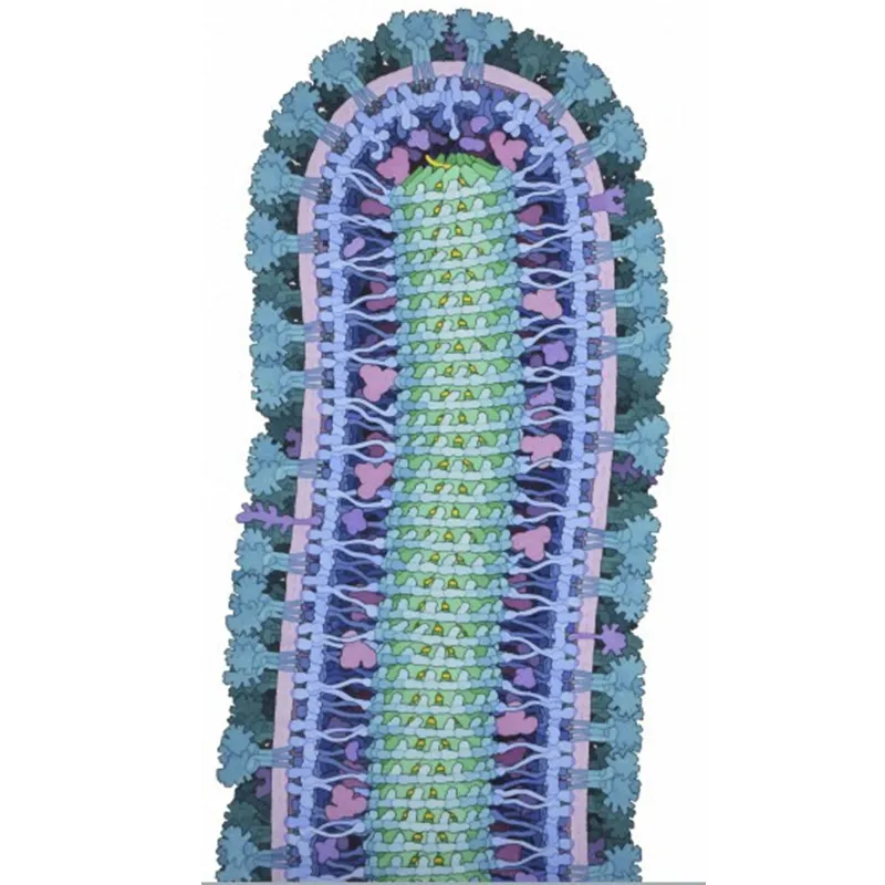
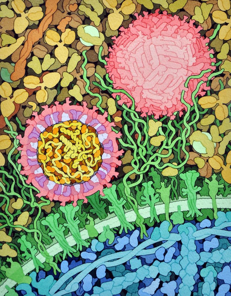
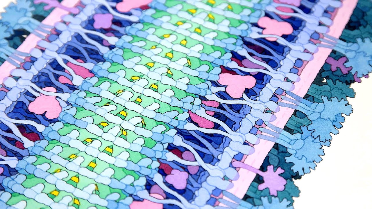
In an Ebola painting, for example, the virus looks like a huge worm rearing its head. The virus has stolen the components of a cell membrane from an infected cell, depicted in light purple, Goodsell writes for the online resource, the RCSB's Protein Data Bank (PDB). Turquoise broccoli-heads stuccoing the outside of that membrane are glycoproteins, which can latch on to the surface of a host cell and pull the viral particle close enough that its genetic material (in yellow, protected by the green nucleoprotein) can be shoved inside. Those glycoproteins have been a major target for drugs to combat the virus.
The painting won this year's Wellcome Image Awards, a competition that draws experts in scientific illustration and visualization from the around the world.
The Ebola painting and many other images by Goodsell live at the PDB, under the supervision of Burley, the repository's director. The PDB holds more than 119,000 structures of proteins, RNA, DNA and other molecules. A few statistics demonstrate how important structure is for biologists: There are about 1.5 million downloads of detailed 3D structural information from the data bank every day. In the last four years, people from 191 of the 194 recognized independent states in the world have accessed the resource.
In July, Goodsell will post his 200th "Molecule of the Month," a series featuring his depictions of proteins and other molecules along with a written explanation of the structures' function and importance.
Goodsell's work helps to educate high school students and others about the structures behind disease-causing particles and health conditions in the news. For the so-called PDB-101 series, his molecules help students better understand the mechanisms behind type 2 diabetes or lead poisoning. He has an upcoming large-scale painting that will cover the life cycle of the HIV virus.
Even the experts can learn from Goodsell's illustrations. Early on, he recalls going around the institute to ask his colleagues how crowded they thought a cell was. The estimates he got back were very dilute. Only when he pulled back to look at the big picture did it become obvious that cells are very dense and complex.
"I'm not aware of many other people operating the way [Goodsell] does," says Burley. Goodsell's work unites artistic interpretation and scientific knowledge. "He is able to tell more of the story of the 3D structure by hand than you can with computer graphics. That, I think, is the real beauty of his work."
Goodsell's work can be seen at the RCSB Protein Data Bank's "Molecule of the Month" series and on his website. His website also provides more detail about some of the images in this article.