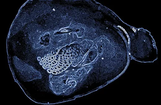
Under the right conditions, patterns emerge from the brain's monumental complexity.
Patric Hagmann (2006) / Abrams Books
Is the human brain, with all its problem-solving prowess and creative ability, powerful enough to understand itself? Nothing in the known universe (with the exception of the universe itself) is more complex; the brain contains about 100 billion nerve cells, or neurons, each of which can communicate with thousands of other brain cells.
Because we primates are primarily visual creatures, perhaps the best way for us to make sense of the brain is to see it clearly. That has been the goal for 125 years, since the Spanish scientist Santiago Ramón y Cajal began using a stain that marked individual neurons. He peered through a microscope at the stained cells and the branchlike projections with which they connected to other neurons. “Here everything was simple, clear and unconfused,” he wrote of his observations, the beginning of modern neuroscience.
Scientists have since devised methods for determining the specific tasks in which different brain regions specialize—for example, some neurons, devoted to processing sight, detect only horizontal lines, while others sense danger or produce speech. Researchers have created maps delineating how brain regions not adjacent to one another are connected by long tracts of cellular projections called axons. The newest microscope techniques reveal neurons changing shape in response to experience—potentially recording a memory. The ability to see the brain in a fresh light has given rise to a wealth of insights in the past few decades.
Now scientists’ forays into this universe are being put to a different use—as art objects. Carl Schoonover, a neuroscientist in training at Columbia University, has collected intriguing images of the brain for a new book, Portraits of the Mind (Abrams). “They are real data, not artists’ renditions,” he says. “This is what neuroscientists are looking at in their microscopes, MRI machines or electrophysiology systems. Neuroscience exists because of these techniques.”
By borrowing a gene from fluorescent jellyfish and inserting it into the DNA of worms or mice in the lab, scientists have made neurons glow. Cajal’s staining technique worked only on post-mortem tissue, and it marked neurons randomly, but the new dyes have enabled scientists to “study neurons in living animals and tissues,” Joshua Sanes of Harvard University notes in an essay in the book.
One of the newest methods relies on a gene that makes algae sensitive to light. Shining a light on neurons containing the gene can change their behavior. “The advances allow us to manipulate the activities of individual cells and cell types using beams of light,” writes Terrence Sejnowski of the Salk Institute for Biological Studies.
The brain remains mysterious, but the patterns in these images—rich whorls of neural connections, unexpected symmetries and layers of structure—encourage scientists to believe they will yet decipher it. For his part, Schoonover hopes to “make readers think it’s worth trying to figure out what the images are and why they are so beautiful.”
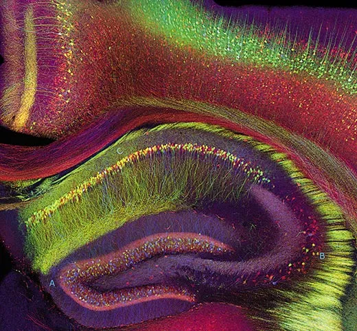
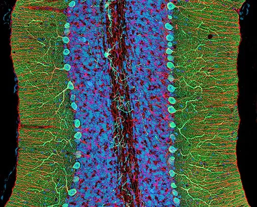
The richly layered hippocampus is where memories are made. The three main components of the hippocampus in this mouse brain are lettered.
Tamily Weissman, Jeff Lichtman and Joshua Sanes (2005) / Abrams Books
/
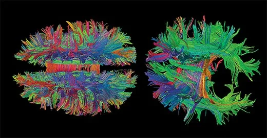
Under the right conditions, patterns emerge from the brain's monumental complexity. One of the newest applications of magnetic resonance imaging tracks the flow of water within cells, revealing neural tracts that make long-distance connections within the brain. In this image of a brain, blue tracts go between the top and bottom, red between right and left, and green between front and back.
Patric Hagmann (2006) / Abrams Books
/
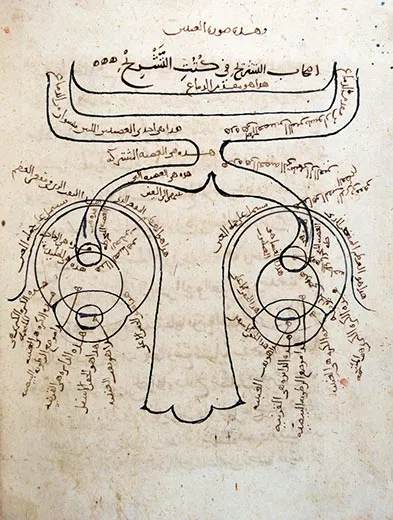
Brain imaging has progressed from gross anatomy to complex circuits. In this first known neuroscience diagram, by Ibn al-Haytham, circa 1027, the eyes and optic nerves are illustrated.
Ibn al-Haytham (circa 1027) / Courtesy of the Süleymaniye Library, Istanbul / Abrams Books
/
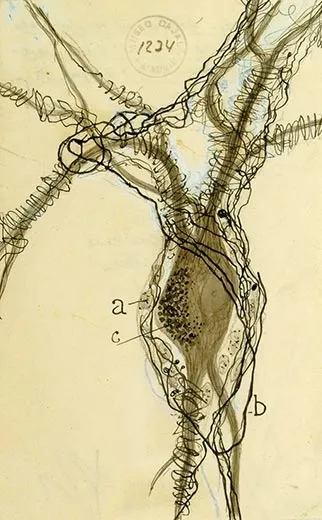
Santiago Ramón y Cajal's 1914 drawing of a plump neuron body entwined by tendrils from other neurons.
Santiago Ramón y Cajal (1914) / Courtesy of Dr. Juan A. de Carlos, Cajal Legacy, Instituto Cajal (CSIC) / Abrams Books
/
The form that a neuron takes is determined by its function, as is the way a group of neurons is organized. Shown here are bright oblong clusters in a part of the mouse brain sensitive to touch; each processes neural signals from a different whisker.
Lasani Wijetunge and Peter Kind, 2008 / Abrams Books
/
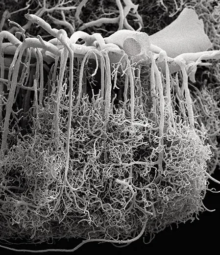
Fueling all this brain activity, and the basis for some imaging techniques, is a dense network of delicate blood vessels.
Alfonso Rodríguez-Baeza and Marisa Ortega-Sánchez (2009) / Abrams Books
/
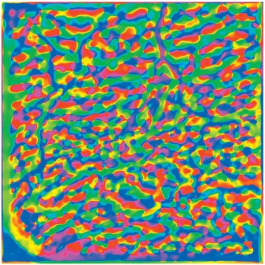
This isn’t abstract art—it’s a representation of neural activity in a monkey’s brain. This part of the brain, called the visual cortex, is one of the first parts of the brain to receive information from the eyes. The visual cortex is tuned to simple shapes, like straight lines. The monkey was shown lines at different orientations, and the different colors represent bits of cortex that are particularly interested in a given type of line. Neuron clusters highlighted in green, for instance, are active when the monkey sees a vertical line; yellow neuron clusters are tuned to horizontal lines.
Courtesy of Yevgeniy B. Sirotin
/
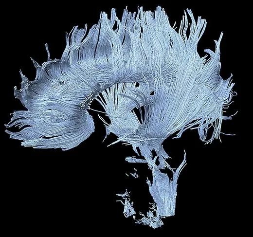
When the brain is working well, the different parts are connected by long fibers called axons (see photo 2). But when the brain is damaged (as in this image from a patient who suffered a stroke in a part of the brain called the thalamus), the connections break down.
Courtesy of Henning U. Voss
/
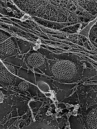
Neurons communicate with one another by releasing chemicals, such as dopamine, from pouches called vesicles. The vesicles, seen here in a fibroblast cell, have a geodesic outer coating that eventually pops through the side of the cell and releases its chemical message to be detected by the cell’s neighbors.
Image produced by John Heuser, MD
/
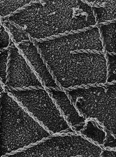
Our cells are surrounded by a scaffold of proteins that maintains a cell’s shape. Under an electron microscope, protein fibers called actin filaments look like braided ropes.
Image produced by John Heuser, MD
/
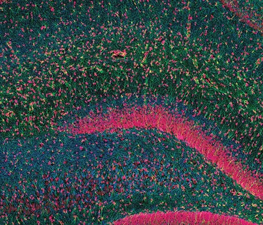
The hippocampus is the seat of memory. If it is damaged, you can remember things that happened long before the injury but you won’t be able to make new memories.
Courtesy of Thomas Deerinck and Mark Ellisman
/
Thank the cerebellum—the convoluted lobe of tissue at the back and bottom of the brain—for your ability to dance or ride a bike. It’s all about motor coordination. In this stained slice of cerebellar tissue, support cells called glia are in blue, and cells called Purkinje neurons are in green. Purkinje neurons are some of the largest neurons in the brain and have extensive branching networks of projections called dendrites.
Courtesy of Thomas Deerinck and Mark Ellisman
/
A few years ago, neuroscientists figured out how to take two fluorescent proteins that glowed in green or red and turn them into a rainbow of different colors that can be incorporated into individual neurons. Here the technique is used to stain cells in the cerebellum. The result? A “brainbow.”
The Brainbow mouse was produced by J. Livet, T.A. Weissman, H. Kang, R.W. Draft, J. Lu, R.A. Bennis, J.R. Sanes, J.W. Lichtman
/
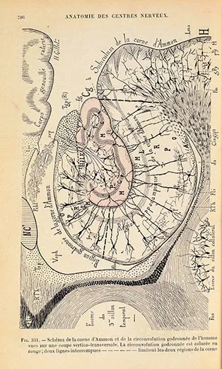
The densely layered hippocampus, which turns out to be crucial for memory, was the subject of this 1895 drawing by Joseph Jules Dejerine.
Photography by Dwight Primiano, Anatomie des centres nerveux. Paris, Rueff, 1895-1901
/
Carl Schoonover’s book includes essays by some of the world’s leading neuroscientists.
Courtesy of Abrams Books
/https://tf-cmsv2-smithsonianmag-media.s3.amazonaws.com/accounts/headshot/laura-helmuth-240.jpg)
/https://tf-cmsv2-smithsonianmag-media.s3.amazonaws.com/filer/brains-magnetic-resonance-imaging-631.jpg)


/https://tf-cmsv2-smithsonianmag-media.s3.amazonaws.com/accounts/headshot/laura-helmuth-240.jpg)