A Mantis Shrimp Inspires a New Camera for Detecting Cancer
The mantis shrimp’s eyes, which can see differences in polarized light, are informing researchers building a tiny, easy-to-use camera that can spot cancer
/https://tf-cmsv2-smithsonianmag-media.s3.amazonaws.com/accounts/headshot/unnamed.jpg)
/https://tf-cmsv2-smithsonianmag-media.s3.amazonaws.com/filer/81/15/81150c66-c97f-41c4-a5cd-1643de798f5f/pseudosquillana_richeri.jpg)
The mantis shrimp is known mostly for its bullet-like punch, which has inspired both super-strong composite materials for future body armor and a viral Web comic about the curious crustacean. But it turns out the animal’s eyes are just as interesting as its claws.
A group of researchers has been working on a way to model the mantis shrimp’s compound eyes and polarized vision to create a camera that can detect various forms of cancer. They now have a proof-of-concept camera sensor that is smaller, simpler and more precise than previous attempts at polarized imaging.
The interdisciplinary group, including a neurobiologist at the University of Queensland, Australia, a computer engineer at Washington University in St. Louis, and others from the University of Maryland, Baltimore County, and the University of Bristol in England, recently published the work in the Proceedings of the IEEE (Institute of Electrical and Electronics Engineers).
The mantis shrimp, like some insects, squid and other cephalopods, can see differences in polarized light—that is light that that is radiating in different planes of direction—in a similar way that we might see the contrast between a black wall and a white table. Animals use this ability to detect prey, find a mate and avoid being eaten.
But polarized light can also be used to see things the human eye can’t, such as cancer cells. The team’s research shows that its sensor has the ability to detect cancerous lesions before the cells become numerous enough to appear as visible tumors.
Viktor Gruev, associate professor of computer science and engineering at Washington University, whose lab worked on building the sensor, says that cancer cells are easy to see under polarized light because their disorganized and invasive structures scatter light differently than normal body cells.
While researchers have created polarized imaging devices in the past, they tend to be large, using multiple sensors, and complex, in that they require optics, engineering and physics experts to operate properly. That, of course, also means the instruments are very expensive.
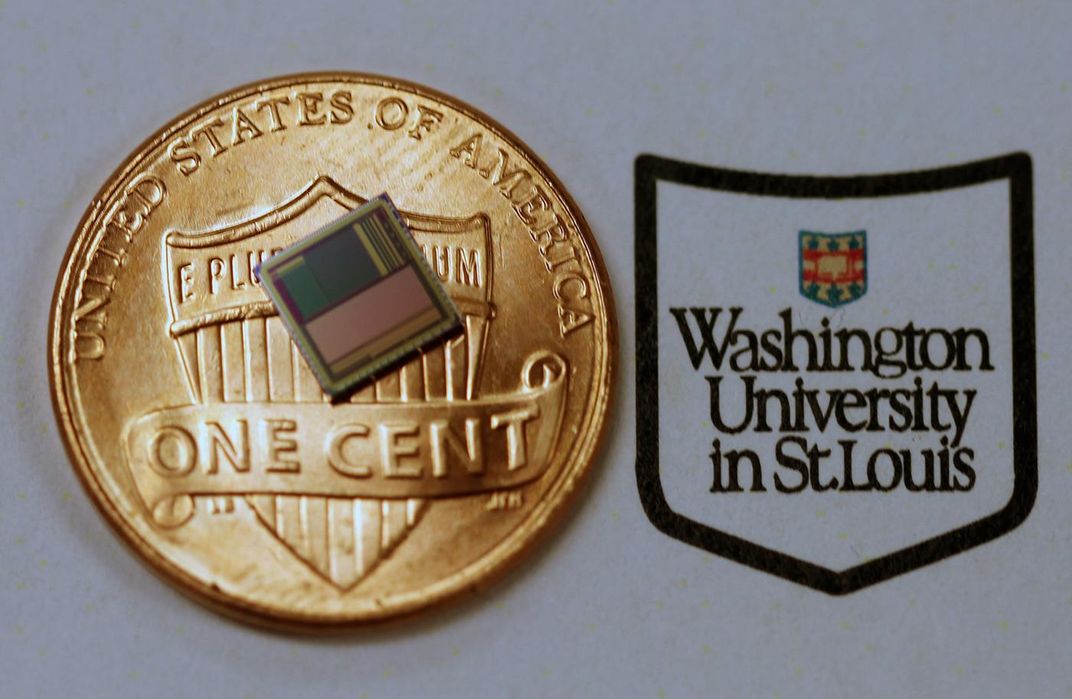
But by combining advancements in nanotechnology, the tiny CMOS (complimentary metal-oxide-semiconductor) sensors common in smartphones and the fundamentals of how the vision system of the mantis shrimp works, the team was able to make a much simpler imaging sensor. Smaller than a penny, the sensor is very sensitive and can detect cancer cells earlier than previous attempts at polarized imaging, using both still images and video. Gruev says his graduate student, Timothy York, the lead author on the paper, did much of the work with the camera and its potential medical applications.
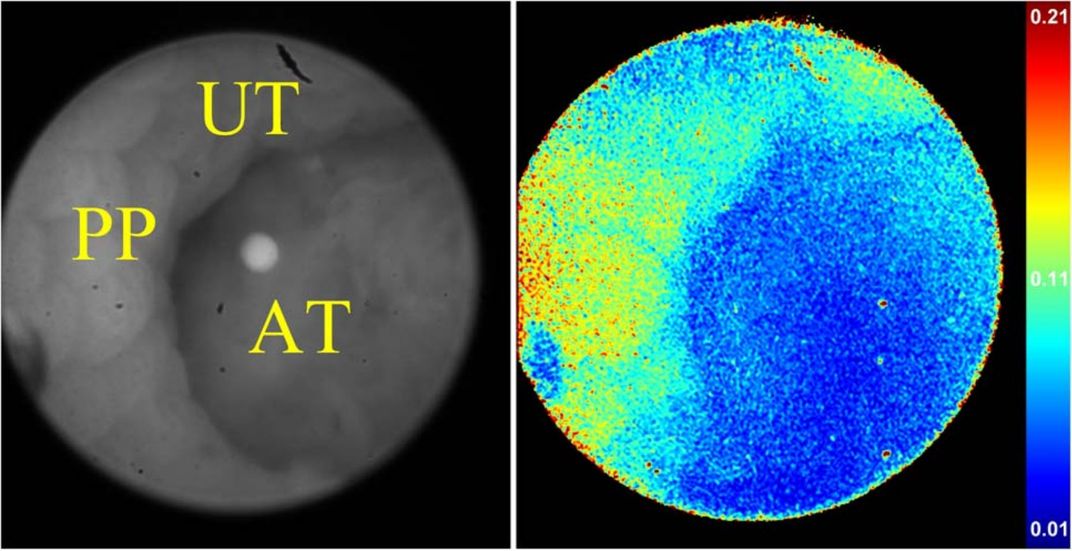
With colon cancer, for instance, a doctor would normally use an endoscope to look for any tissue that looks cancerous, then take a biopsy. But the cancer has to be at a certain stage of development before it looks different to the human eye. Polarized imaging can spot cancer cells much earlier, but previous imaging devices have been too large to be used in this way before.
“We’ve moved from having multiple cameras to a single-chip solution,” says Gruev. “It’s hard to put multiple cameras on an endoscope and take pictures. With our device, all the filters are on the camera and it goes from something that sits on your optical bench to one that goes on the end of an endoscope.”
The camera could drastically reduce the need for biopsies—but until the technology is refined, the extent to which it will do so is unclear.
Justin Marshall, a neurobiologist at the University of Queensland and another of the paper’s authors, brought his expertise on mantis shrimp to the project. He has been investigating the shrimp's vision for more than 25 years. Both he and Gruev agree that one of the next challenges will be to find a way to incorporate traditional color vision into the sensor as well. As it stands now, the sensor can see differences in polarization, but not the colors that we see. That is a problem for doctors who may one day use this type of sensor, because they typically use visual cues to guide them during delicate procedures. But shrimp could provide some help on that front as well.
“[Mantis shrimp] seem to be very particular about the way they gather information, both in terms of color and polarization,” says Marshall. “They wave their eyes around in order to push their sensor over the world, a bit like a satellite scanning. There may be some tricks in there that we can borrow from as well.”
Marshall thinks the sensor may be used to screen patients for colon cancer first, as that's a specific area his team has been working on and one where the size and complexity of other polarized imaging cameras has been a problem in the past. Simpler polarization scopes are already being used to check for skin cancer in Australia, where two in three people are diagnosed with the disease before the age of 70. The researchers are also experimenting with using polarized light to increase tissue contrast to help doctors tell where to start and stop cutting during surgery.
Because the shrimp-inspired chip is so compact and easy to use, the technology could make its way into portable devices and even smartphones. If it does, Marshall says, people could one day self-monitor for cancers and reduce the burden on overloaded healthcare systems.
While there is plenty of potential in the polarized imaging technology, Gruev says there is still a lot of work to be done, both in incorporating color sensing and in refining the sensitivity of the polarization detection to increase resolution and make it even better at detecting serious illnesses early.
“We are just scratching the surface of how we can look at biology and construct imaging systems that can help in the diagnosis of cancer and other diseases,” he says.
/https://tf-cmsv2-smithsonianmag-media.s3.amazonaws.com/accounts/headshot/unnamed.jpg)